

Surgical approach to the treatment of pituicytoma. Report of five cases and a literature review
- PMID: 37026087
- PMCID: PMC10070180
- DOI: 10.1016/j.wnsx.2023.100186
Surgical approach to the treatment of pituicytoma. Report of five cases and a literature review
Abstract
Background: Pituicytoma (PTs) is a rare tumor of the sella and suprasellar region, derived from the pituicytes of the neurohypophysis, having distinct histological characteristics of glial neoplasms. We reported, the clinical data, neuroimaging studies, surgical approaches and pathology in five patients with PTs and also, we reviewed the literature.
Methods: Retrospective chart from five consecutive patients with PTs treated at one University Hospital from 2016 to 2021 were reviewed. In addition, we conducted a search in PubMed/Medline databases using the term "Pituicytoma". Data regarding age, gender, pathological findings, and treatment modality applied were extracted.
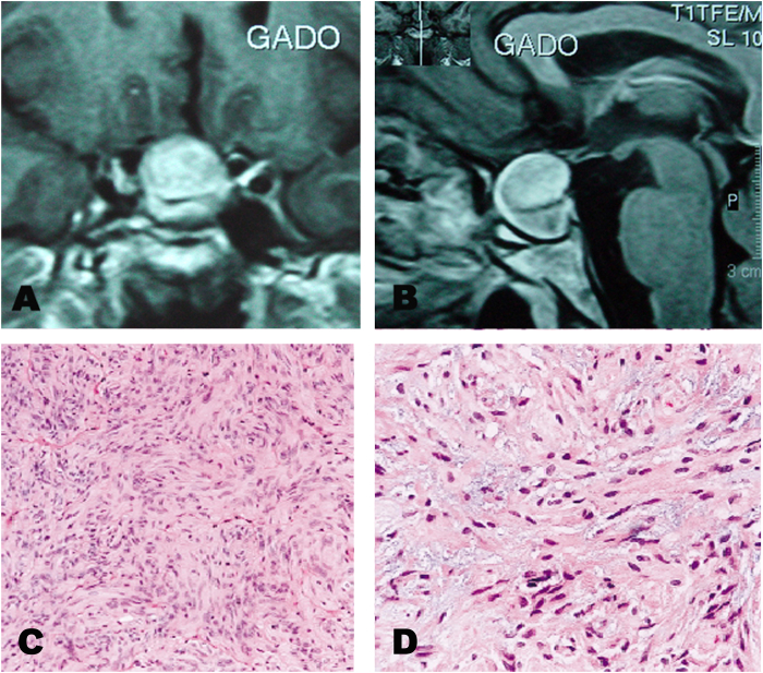
Results: All patients were female, aged 29-63, complaining of headaches, visual loss and field defects, dizziness and normal or abnormal levels of circulating pituitary hormones. Magnetic Resonance Imaging (MRI) showed in all patients a sellar and suprasellar mass, which was removed through an endoscopic transsphenoidal approach. Our third patient had a subtotal resection followed by close observation. Histopathology showed a glial non-infiltrative tumors with spindle cells, and a final diagnosis of pituicytoma was made. After surgery, visual field defects in all patients were normalized, and in two patients normal levels of plasma hormones were restored. After a mean of three years follow-up, the patients were managed post-operatively through close clinical observation and serial MRI. None of the patients had recurrence of the disease.
Conclusion: PTs is a rare glial tumor of the sellar and suprasellar region that arises from neurohypophyseal pituicytes. Disease control may be achieved by total excision.
Keywords: Coronavirus; Glioma; Neurohypophyseal disease; Pituitary tumor.
© 2023 The Authors.
Conflict of interest statement
The authors of “Surgical approach to treatment of pituicytoma. Criteria applied in a patient having COVID-19 and literature review.” declare that they have no relevant or material financial interests that relate to the research described in this paper.
Figures


References
-
- Brat D.J., Scheithauer B.W., Staugaitis S.M., Holtzman R.N., Morgello S., Burger P.C. Pituicytoma: a distinctive low-grade glioma of the neurohypophysis. Am J Surg Pathol. 2000;24:362–368. - PubMed
-
- Cenacchi G., Giovenali P., Castrioto C., Giangaspero F. Pituicytoma: ultrastructural evidence of a possible origin from folliculo-stellate cells of the adenohypophysis. Ultrastruct Pathol. 2001;25:309–312. - PubMed
-
- Feiden S., Feiden W. WHO classification of tumors of the CNS: revised edition of 2007 with critical comments on the typing of common-type diffuse gliomas. Pathologe. 2008;29(6):411–421. - PubMed
-
- Katsuta T., Inoue T., Nakagaki H., Takeshita M., Morimoto K., Iwaki T. Distinctions between pituicytoma and ordinary pilocytic astrocytoma. Case report. J Neurosurg. 2003;98:404–406. - PubMed
-
- Nakazato Y. Revised WHO classification of brain tumors. Brain Nerve. 2008;60(1):59–77. - PubMed
LinkOut - more resources
Full Text Sources
