

Lacrimal and meibomian gland evaluation in dry eye disease: A mini-review
- PMID: 37026239
- PMCID: PMC10276709
- DOI: 10.4103/IJO.IJO_2622_22
Lacrimal and meibomian gland evaluation in dry eye disease: A mini-review
Abstract
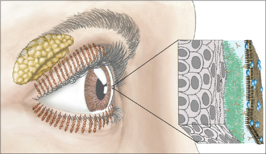
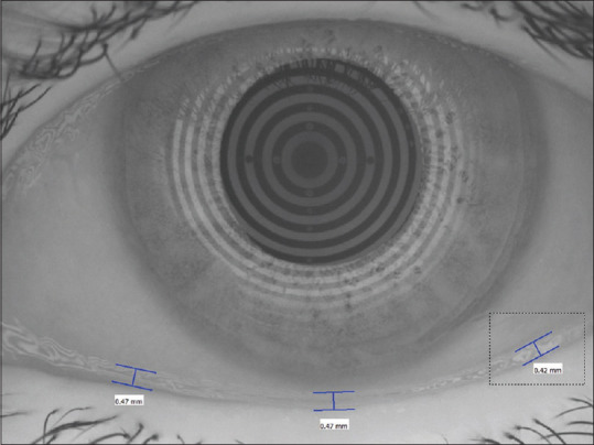
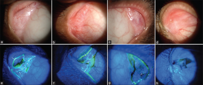
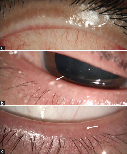
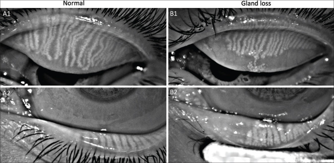
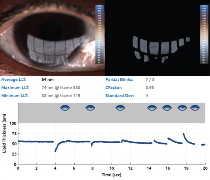
Lacrimal and meibomian glands contribute to the aqueous and lipid components of tear film, respectively. Their evaluation remains central to diagnosing and managing dry eye disease (DED). The review discusses the differences and reliability of various diagnostic tests and commercially available devices used for DED diagnosis. Slit-lamp-based techniques are direct palpebral lobe and tear flow assessment, Schirmer test, meibum quality and expressibility, and evaluation of tear meniscus height. Non-invasive tear meniscus height (TMH), tear break-up time (TBUT), lipid layer thickness (LLT), and meibography are machine-based diagnostic tests. The structure-function correlation of the tear-producing glands gives more comprehensive details than either information alone. Many devices are available in the market, which make DED diagnosis an easy feat, but the tests should be interpreted keeping in mind the intra-observer and inter-observer repeatability. Also, the tear film displays a huge variability as per the environmental conditions and impact of blinking. Hence, the examiner should be well versed with the techniques and repeat the test two to three times to obtain an average reading, which is more reliable. The recommended sequence of tests for diagnosing DED is a dry eye questionnaire, TMH, LLT, NIBUT (FBUT if non-invasive test is unavailable but should be performed after osmolarity), tear osmolarity, meibography, and ocular surface staining. Invasive tests such as Schirmer should be performed after the non-invasive tear film diagnostic testing.
Keywords: Dry eye disease; lacrimal gland; lipid layer thickness; meibomian glands; tear break-up time; tear meniscus height.
Conflict of interest statement
None
Figures

References
-
- Singh S, Basu S. The lacrimal gland:Historical perspectives and current understanding. Curr Eye Res. 2020;45:1188–98. - PubMed
-
- Singh S, Basu S. Secretory ductules of lacrimal gland. Ophthalmic Plast Reconstr Surg. 2021;37:e83. - PubMed
-
- Singh S, Shanbhag SS, Basu S. Palpebral lobe of human lacrimal gland:Morphometric analysis in normal versus dry eyes. Br J Ophthalmol. 2021;105:1352–7. - PubMed
-
- Singh S, Das AV, Basu S. Ocular involvement in Sjogren's syndrome:Risk factors for severe visual impairment and vision threatening corneal complications. Am J Ophthalmol. 2021;225:11–7. - PubMed
-
- Singh S, Ali MJ, Mittal V, Brabletz S, Paulsen F. Immunohistological study of palpebral lobe of the lacrimal gland in severe dry eyes secondary to Stevens-Johnson syndrome. Curr Eye Res. 2021;46:789–95. - PubMed
Publication types
MeSH terms
Substances
LinkOut - more resources
Full Text Sources
Medical
