

Keratoprosthesis in dry eye disease
- PMID: 37026247
- PMCID: PMC10276669
- DOI: 10.4103/IJO.IJO_2817_22
Keratoprosthesis in dry eye disease
Abstract
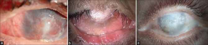
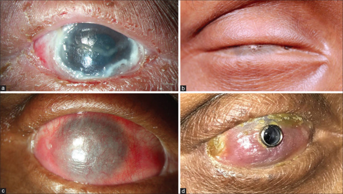
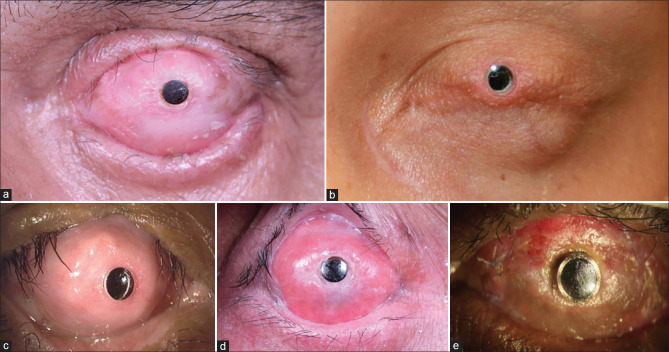
Bilateral corneal blindness with severe dry eye disease (DED), total limbal stem cell deficiency with underlying corneal stromal scarring and vascularization, combined with adnexal complications secondary to chronic cicatrizing conjunctivitis is a highly complex situation to treat. In such eyes, procedures such as penetrating keratoplasty alone or combined with limbal stem cell transplantation are doomed to fail. In these eyes, keratoprosthesis (Kpro) or an artificial cornea is the most viable option, eliminating corneal blindness even in eyes with autoimmune disorders such as Stevens-Johnson syndrome, ocular mucous membrane pemphigoid, Sjogren's syndrome, and nonautoimmune disorders such as chemical/thermal ocular burns, all of which are complex pathologies. Performing a Kpro in these eyes also eliminates the need for systemic immunosuppression and may provide relatively early visual recovery. In such eyes, the donor cornea around the central cylinder of the Kpro needs to be covered with a second layer of protection to avoid desiccation and progressive stromal melt of the underlying cornea, which is a common complication in eyes with severe DED. In this review, we will focus on Kpro designs that have been developed to survive in eyes with the hostile environment of severe DED. Their outcomes in such eyes will be discussed.
Keywords: Chronic cicatrizing conjunctivitis; Stevens–Johnson syndrome; dry eye disease; keratoprosthesis; limbal stem cell deficiency; mucous membrane pemphigoid; ocular chemical burns.
Conflict of interest statement
None
Figures
Comment in
-
Comment on "Keratoprosthesis in dry eye disease".Indian J Ophthalmol. 2023 Sep;71(9):3267. doi: 10.4103/IJO.IJO_940_23. Indian J Ophthalmol. 2023. PMID: 37602627 Free PMC article. No abstract available.
References
-
- Chirila TV, Hicks CR. The origins of the artificial cornea:Pellier de Quengsy and his contribution to the modern concept of keratoprosthesis. Gesnerus. 1999;56:96–106. - PubMed
-
- Saeed HN, Shanbhag S, Chodosh J. The Boston keratoprosthesis. Curr Opin Ophthalmol. 2017;28:390–6. - PubMed
-
- Pujari S, Siddique SS, Dohlman CH, Chodosh J. The Boston keratoprosthesis Type II:The Massachusetts eye and ear infirmary experience. Cornea. 2011;30:1298–303. - PubMed
-
- Alexander JK, Basak SK, Padilla MD, Yu F, Aldave AJ. International outcomes of the Boston Type I Keratoprosthesis in Stevens–Johnson syndrome. Cornea. 2015;34:1387–94. - PubMed
Publication types
MeSH terms
LinkOut - more resources
Full Text Sources
Medical
