

Tear biomarkers in dry eye disease: Progress in the last decade
- PMID: 37026250
- PMCID: PMC10276712
- DOI: 10.4103/IJO.IJO_2981_22
Tear biomarkers in dry eye disease: Progress in the last decade
Abstract
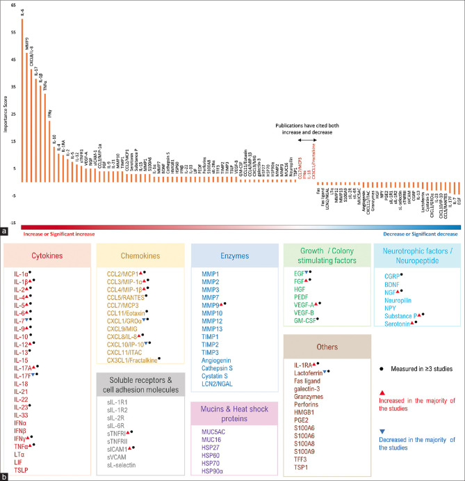
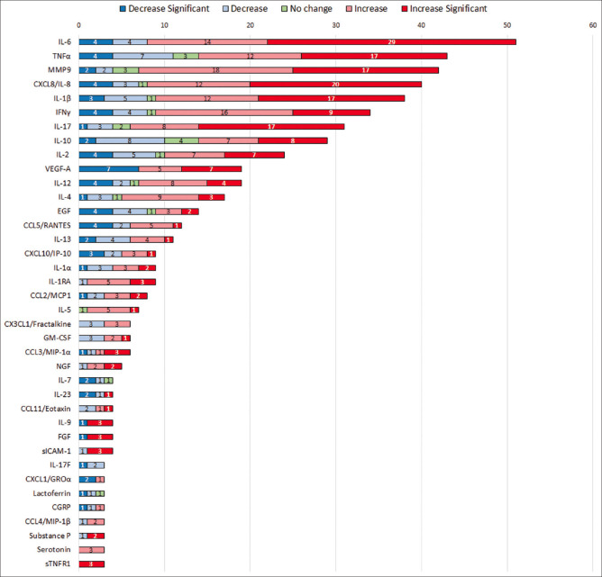
Dry eye disease (DED) is a commonly occurring, multifactorial disease characterized by reduced tear film stability and hyperosmolarity at the ocular surface, leading to discomfort and visual compromise. DED is driven by chronic inflammation and its pathogenesis involves multiple ocular surface structures such as the cornea, conjunctiva, lacrimal glands, and meibomian glands. The tear film secretion and its composition are regulated by the ocular surface in orchestration with the environment and bodily cues. Thus, any dysregulation in ocular surface homeostasis causes an increase in tear break-up time (TBUT), osmolarity changes, and reduction in tear film volume, all of which are indicators of DED. Tear film abnormalities are perpetuated by underlying inflammatory signaling and secretion of inflammatory factors, leading to the recruitment of immune cells and clinical pathology. Tear-soluble factors such as cytokines and chemokines are the best surrogate markers of disease severity and can also drive the altered profile of ocular surface cells contributing to the disease. Soluble factors can thus help in disease classification and planning treatment strategies. Our analysis suggests increased levels of cytokines namely interleukin-1β (IL-1β), IL-2, IL-4, IL-6, IL-9, IL-12, IL-17A, interferon-gamma (IFN-γ), tumor necrosis factor-alpha (TNF-α); chemokines (CCL2, CCL3, CCL4, CXCL8); MMP-9, FGF, VEGF-A; soluble receptors (sICAM-1, sTNFR1), neurotrophic factors (NGF, substance P, serotonin) and IL1RA and reduced levels of IL-7, IL-17F, CXCL1, CXCL10, EGF and lactoferrin in DED. Due to the non-invasive sample collection and ease of quantitively measuring soluble factors, tears are one of the best-studied biological samples to molecularly stratify DED patients and monitor their response to therapy. In this review, we evaluate and summarize the soluble factors profiles in DED patients from the studies conducted over the past decade and across various patient groups and etiologies. The use of biomarker testing in clinical settings will aid in the advancement of personalized medicine and represents the next step in managing DED.
Keywords: Biomarker; chemokines; cytokines; dry eye disease; growth factors; tear-soluble factors.
Conflict of interest statement
None
Figures


References
-
- Craig JP, Nichols KK, Akpek EK, Caffery B, Dua HS, Joo CK, et al. TFOS DEWS II definition and classification report. Ocul Surf. 2017;15:276–83. - PubMed
-
- Cai Y, Wei J, Zhou J, Zou W. Prevalence and incidence of dry eye disease in Asia:A systematic review and meta-analysis. Ophthalmic Res. 2022;65:647–58. - PubMed
-
- Donthineni PR, Kammari P, Shanbhag SS, Singh V, Das AV, Basu S. Incidence, demographics, types and risk factors of dry eye disease in India:Electronic medical records driven big data analytics report I. Ocul Surf. 2019;17:250–6. - PubMed
Publication types
MeSH terms
Substances
LinkOut - more resources
Full Text Sources
Miscellaneous
