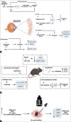
Autophagy in dry eye disease: Therapeutic implications of autophagy modulators on the ocular surface
- PMID: 37026260
- PMCID: PMC10276742
- DOI: 10.4103/IJO.IJO_2912_22
Autophagy in dry eye disease: Therapeutic implications of autophagy modulators on the ocular surface
Abstract
Dry eye disease (DED) is a chronic ocular surface disorder, associated with inflammation, which can cause severe morbidity, visual compromise, and loss of quality of life, affecting up to 5-50% of the world population. In DED, ocular surface damage and tear film instability due to abnormal tear secretion lead to ocular surface pain, discomfort, and epithelial barrier disruption. Studies have shown the involvement of autophagy regulation in dry eye disease as a pathogenic mechanism along with the inflammatory response. Autophagy is a self-degradation pathway in mammalian cells that reduces the excessive inflammation driven by the secretion of inflammatory factors in tears. Specific autophagy modulators are already available for the management of DED currently. However, growing studies on autophagy regulation in DED might further encourage the development of autophagy modulating drugs that reduce the pathological response at the ocular surface. In this review, we summarize the role of autophagy in the pathogenesis of dry eye disease and explore its therapeutic application.
Keywords: Autophagy modulators; corneal autophagy; inflammation; ocular surface dry eye disease.
Conflict of interest statement
None
Figures


References
-
- Craig JP, Nichols KK, Akpek EK, Caffery B, Dua HS, Joo CK, et al. TFOS DEWS II definition and classification report. Ocul Surf. 2017;15:276–83. - PubMed
-
- Wolffsohn JS, Arita R, Chalmers R, Djalilian A, Dogru M, Dumbleton K, et al. TFOS DEWS II diagnostic methodology report. Ocul Surf. 2017;15:539–574. - PubMed
-
- Doughty MJ. Rose bengal staining as an assessment of ocular surface damage and recovery in dry eye disease-a review. Cont Lens Anterior Eye. 2013;36:272–80. - PubMed
-
- Jones L, Downie LE, Korb D, Benitez-Del-Castillo JM, Dana R, Deng SX, et al. TFOS DEWS II management and therapy report. Ocul Surf. 2017;15:575–628. - PubMed
Publication types
MeSH terms
LinkOut - more resources
Full Text Sources

