Review
. 2023 Jun;10(16):e2206939.
doi: 10.1002/advs.202206939. Epub 2023 Apr 7.

Spatial Transcriptomics: Technical Aspects of Recent Developments and Their Applications in Neuroscience and Cancer Research

Affiliations
Review

Spatial Transcriptomics: Technical Aspects of Recent Developments and Their Applications in Neuroscience and Cancer Research

Han-Eol Park et al. Adv Sci (Weinh). 2023 Jun.

Abstract

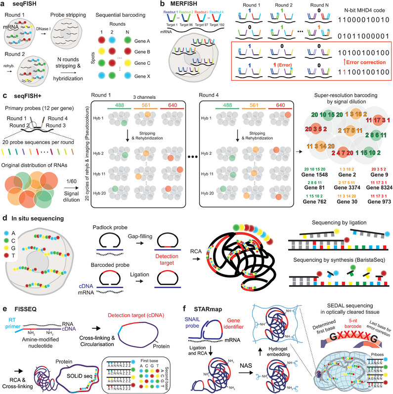
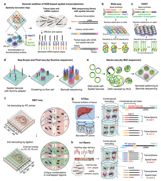
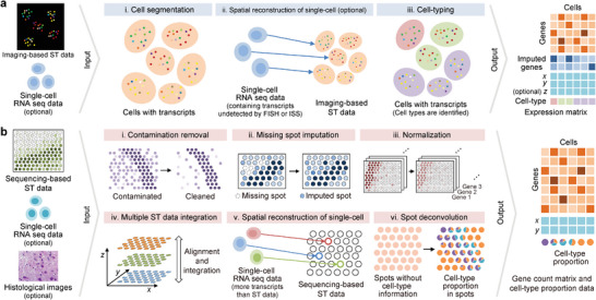
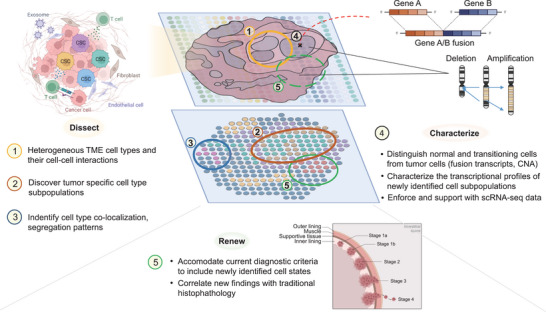
Spatial transcriptomics is a newly emerging field that enables high-throughput investigation of the spatial localization of transcripts and related analyses in various applications for biological systems. By transitioning from conventional biological studies to "in situ" biology, spatial transcriptomics can provide transcriptome-scale spatial information. Currently, the ability to simultaneously characterize gene expression profiles of cells and relevant cellular environment is a paradigm shift for biological studies. In this review, recent progress in spatial transcriptomics and its applications in neuroscience and cancer studies are highlighted. Technical aspects of existing technologies and future directions of new developments (as of March 2023), computational analysis of spatial transcriptome data, application notes in neuroscience and cancer studies, and discussions regarding future directions of spatial multi-omics and their expanding roles in biomedical applications are emphasized.

Keywords: RNA; bioinformatics; cancer; neuroscience; spatial transcriptomics.


Conflict of interest statement

The authors declare no conflict of interest.

Figures

Figure 1
Figure 1
Imaging‐based spatial transcriptomics methods. a) seqFISH by DNaseI‐based digestion and sequential staining/imaging cycles to decode transcripts in space. b) MERFISH employing error correction in barcode assignment for robust barcode calling in noisy FISH‐based images. c) seqFISH+ for genome‐scale transcriptome investigation by dilution of fluorescent signals, separating individual transcripts into fluorescent spectra, and employing 20 probes per each encoding round. d) in situ sequencing methods by sequencing by ligation (ISS, FISSEQ, STARmap) and sequencing by synthesis (BaristaSeq). e) SOLiD sequencing of cDNA sequence by FISSEQ while cross‐linking cDNA and amplicon generated by rolling‐circle amplification (RCA) to adjacent proteins. f) STARmap with SNAIL probes and SEDAL sequencing for identifying gene‐specific identifiers. The polymerization of amplicons with acrylamide moieties introduced by N‐acryloxysuccinimide (NAS) into the hydrogel network and optical clearing by hydrogel‐histochemistry enables spatial transcriptome detection in thick tissues.
Figure 2
Figure 2
Next‐generation sequencing (NGS)‐based spatial transcriptomics methods. a) General workflow of the spatial transcriptomics (ST) method. The spatial barcode‐encoded oligos are immobilized on a functionalized surface to capture mRNA released from the mounted tissue or cells. Subsequent cDNA synthesis, followed by sequencing libraries yield transcript sequences and their spatial locations, simultaneously. b–e) Developments of methods for the spatial patterning of barcoded oligos with enhanced spatial resolution. Unlike imaging‐based ST, these methods require barcode decoding after patterning due to the random spatial distribution of spatial barcodes. b) Slide‐seq employs random spatial bead spreading and in situ sequencing decoding. c) HDST deposits beads with combinatorial barcodes on patterned wafers, followed by decoding with serial hybridization. d) Seq‐Scope and Pixel‐seq utilize Illumina clustering for oligo patterning and directly read sequences using Illumina sequencers. e) Stereo‐seq utilizes DNBSEQ chemistry to generate DNA nanoballs with spatial barcodes, which are patterned on a flow cell, and barcode calling is performed by the MGI sequencer. f) DBiT‐seq delivers barcoded RT primers and ligation oligos through orthogonal microfluidic channels. The predetermined spatial distribution of overlapping regions eliminates time‐consuming steps for random spatial barcode sequencing procedures. g) True single‐cell and single‐nucleus resolutions with regional spatial barcode printing in XYZeq. h) sci‐Space delivers hashing oligos with spatial barcodes into tissue followed by additional fixation to retain hashing oligos in the nuclei. After combinatorial barcoding for single‐nucleus RNA sequencing, hashing oligos are sequenced as a transcriptome.
Figure 3
Figure 3
The workflows for preprocessing raw data in a) imaging‐based and b) sequencing‐based spatial transcriptomics methods. In the imaging‐based workflow, spatial transcriptomics data is predominantly analyzed independently, while single‐cell sequencing data is occasionally integrated. In contrast, sequencing‐based methods more commonly employ both single‐cell sequencing data and histological images simultaneously. The resulting outputs of the two workflows differ in their celltype format. For sequencing‐based methods, the spot size is usually larger than a single cell, so the cell type of each spot is described proportionally. In contrast, imaging‐based methods provide a cell‐level gene count matrix, which directly labels the cell type on each cell. These outputs are then employed for downstream analysis.
Figure 4
Figure 4
Applications of spatial transcriptomics in neuroscience. The transcriptomic atlas is established by cell‐type identification with the spatial context of precisely compartmentalized brain structure. Transcriptome‐based neuronal cell types can be further dissected by integrative studies employing tools for functionality and connectivity investigations. Accurate neural classification will lead to circuit‐level studies to investigate individual circuits to elucidate the comprehensive function of the entire brain.
Figure 5
Figure 5
Applications of spatial transcriptomics in cancer research. Spatially resolved transcriptomics can be used to define the cell type compositions and discover new cell–cell interactions of specific tumor ecosystems, profile and characterize these ecosystems by utilization and integration in multi‐modal/omics analyses, and help fuel the joint renewal of current histopathological standards to accommodate these new findings. (Figure created with icons and redesigned templates provided by Biorender.com).

References

    1. Lewis J., Science 2008, 322, 399. - PubMed
    1. Lander A. D., Science 2013, 339, 923. - PMC - PubMed
    1. Cremisi F., Philpott A., Ohnuma S., Curr. Opin. Neurobiol. 2003, 13, 26. - PubMed
    1. Fuchs E., Tumbar T., Guasch G., Cell 2004, 116, 769. - PubMed
    1. Rompolas P., Mesa K. R., Greco V., Nature 2013, 502, 513. - PMC - PubMed

Publication types

LinkOut - more resources