. 2023 Jun 5;222(6):e202209098.
doi: 10.1083/jcb.202209098. Epub 2023 Apr 7.

Toxoplasma ERK7 protects the apical complex from premature degradation

Affiliations

Toxoplasma ERK7 protects the apical complex from premature degradation

William J O'Shaughnessy et al. J Cell Biol. .

Abstract

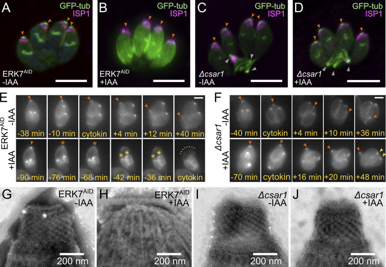
Accurate cellular replication balances the biogenesis and turnover of complex structures. In the apicomplexan parasite Toxoplasma gondii, daughter cells form within an intact mother cell, creating additional challenges to ensuring fidelity of division. The apical complex is critical to parasite infectivity and consists of apical secretory organelles and specialized cytoskeletal structures. We previously identified the kinase ERK7 as required for maturation of the apical complex in Toxoplasma. Here, we define the Toxoplasma ERK7 interactome, including a putative E3 ligase, CSAR1. Genetic disruption of CSAR1 fully suppresses loss of the apical complex upon ERK7 knockdown. Furthermore, we show that CSAR1 is normally responsible for turnover of maternal cytoskeleton during cytokinesis, and that its aberrant function is driven by mislocalization from the parasite residual body to the apical complex. These data identify a protein homeostasis pathway critical for Toxoplasma replication and fitness and suggest an unappreciated role for the parasite residual body in compartmentalizing processes that threaten the fidelity of parasite development.


Conflict of interest statement

Disclosures: The authors declare no competing interests exist.

Figures

Figure 1.
Figure 1.
Toxoplasma divides by endodyogeny. (A) In normal Toxoplasma division, two daughter parasites are formed within the intact mother cell and are delineated by a specialized cytoskeleton and membrane structure called the inner membrane complex (IMC; green). During cytokinesis, the daughter cells integrate their cytoskeleton into the mother plasma membrane. After division, the parasites remain connected through the “residual body.” (B) Diagram of ERK7 kinase loss-of-function phenotype in Toxoplasma division. When parasites divide without functional ERK7 kinase localized at their apical tips, the conoid (magenta) is lost in the mature parasite after cytokinesis, rendering the parasites noninvasive. Note that new daughter parasites grow and develop with a visible conoid.
Figure S1.
Figure S1.
Candidates from the ERK7 interactome were examined by creating screens in which the gene of interest was expressed in-frame with a C-terminal 3xHA tag in the background of the ERK7AID strain. Parasites were grown in ±IAA for 24 h before fixation. Samples were stained with antibodies recognizing the HA-tag (green), tubulin (blue), and the indicated counter-stain (red; usually ISP1, a marker for the apical cap; in some cases IMC1 a marker for the inner-membrane complex; ROP2 a marker for the rhoptries; MIC2 a marker for the micronemes; GRA2 a marker for secretion into the parasitophorous vacuole). For unmerged images, see preprint (O’Shaughnessy et al., 2023). All images are representative of a minimum of n = 20 images and are maximum intensity Z-projections of confocal stacks. While TGGT1_236560 was a hit in both our Yeast-two-hybrid and BioID datasets, C-terminal tagging led to vacuole localization, which is inconsistent with an interaction with cytosolic ERK7. TGGT1_236560 also has no predicted signal peptide. Note that the predicted protein length is ∼250 kD, and the gene has no predicted introns, which is unusual for a gene of this size. It is therefore possible that the gene model is incorrect and represents a combination of a cytosolic protein (at the 5′ end of the gene) and a secreted protein (at the 3′ end).
Figure 2.
Figure 2.
CSAR1 mislocalizes to daughter buds upon ERK7 degradation. (A and B) Maximum intensity Z-projections of confocal stacks of CSAR1-3xHA ERK7AID parasites grown for 24 h (A) without IAA and (B) with IAA to degrade ERK7AID. Parasites were visualized with GFP-tubulin (blue), and antibodies against HA (green) and the apical cap marker ISP1 (red). Concentration of CSAR1 signal at the residual body is indicated with a white arrow. The apical ends of the late daughter buds in the + IAA condition are indicated with orange arrows. Parasites were captured at the indicated points in the cell cycle. (C) Maximum intensity Z-projects of confocal stack of CSAR1-3XHA ERK7AID parasites counterstained with an anti-ERK7 antibody. All scale bars are 10 μm.
Figure S2.
Figure S2.
CSAR1 exhibits dynamic localization through the Toxoplasma cell cycle. (A) Late in mitosis (monitored by GFP-tubulin signal; green), in 68% of vacuoles examined, CSAR13xHA signal (magenta) is bright and confined to the maternal cytosol (see Fig. S3, below). In 32% of these vacuoles, CSAR13xHA has begun to concentrate at the residual body (lower panel; yellow arrowhead). (B and C) As the vacuoles with CSAR1-residual body signal tend to contain larger daughter buds on average (B), we assume this relocalization of CSAR1 is occurring in parasites that have progressed further in mitosis, and which we therefore refer to in C as “Very late mitosis.” P value is from 2-tailed unpaired Student’s t test. Normality tested by Shapiro-Wilk test. (C) Gallery of representative images of CSAR13xHA localization during cell cycle in parasites untreated with IAA during the second division cycle (2 → 4 parasites).
Figure S3.
Figure S3.
CSAR1 is excluded from daughter buds. (A) 0.5 μm slice through a budding CSAR13xHA parasites from a confocal stack. CSAR13xHA (magenta), while building up in the maternal cytosol, appears excluded from the cytosol of daughter buds, which are outlined by GFP-tubulin (green). Scale bar is 5 μm. (B) A line scan at the indicated position in A highlights the lack of CSAR13xHA signal within a daughter bud.
Figure 3.
Figure 3.
CSAR1 is required for full efficiency of lytic cycle. (A) Western blot stained with anti-FLAG (recognizing ERK7AID-3xFLAG) and anti-tubulin of lysates from ERK7AID and Δcsar1 parasites grown in ±IAA. (B–F) Quantification of (B) plaque number, (C) relative plaque size, (D) invasion rate, (E) ionophore-induced egress, and (F) replication rates of ERK7AID or Δcsar1 parasites grown in ±IAA binned as indicated. B and C also show relative fitness of the wild-type 3xHA-tagged and RING-domain mutant CSAR1 (C1791A/H1792A/H1796A; CSAR1MUT) parasites. Significance on B–E calculated by 1-way ANOVA followed by Tukey’s multiple comparison test (*** is P < 0.001; n.s., not significant). Data distribution was assumed to be normal, but this was not formally tested. (G) The unbinned cumulative frequency of the data from F. Invasion, egress, and plaque assays are n = 3 biological replicates conducted with n = 3 technical replicates. Replication quantified from three biological replicates of n ≥ 100 vacuoles. Error bars in F are SD, all others are 95% confidence-interval of mean. Replication of Δcsar1 compared to parental ERK7AID is P < 0.001 as calculated by Kolmogorov-Smirnov test. Source data are available for this figure: SourceData F3.
Figure 4.
Figure 4.
Loss of CSAR1 causes retention of maternal cytoskeleton in the residual body. (A and B) Maximum intensity Z-projections of confocal stacks of (A) ERK7AID and (B) Δcsar1 parasites grown in the absence of IAA. Parasites were visualized with GFP-tubulin (green), and antibodies against the inner membrane complex marker IMC1 (red) and ERK7 (blue). Apical ends of parasites are indicated with white arrows. Retained cytoskeleton from the previous two divisions in the Δcsar1 vacuole is indicated with orange arrows. Scale bars are 5 μm.
Figure 5.
Figure 5.
Retained maternal cytoskeleton is contained in membrane of the residual body. (A) Correlated fluorescence (GFP-tub; inset scale bar, 5 μm) and transmission electron microscopy of Δcsar1-infected fibroblasts. Boxed regions are zoomed in the indicated panels. (B and C) Structures consistent with parasite microtubule cytoskeleton are found within the residual body. (B′ and B″) serial slices of the region in B show multiple retained conoids are tightly packed within the residual body. (D) Cross-section of a bundle of 20 subpellicular microtubules delineated by the residual body membrane. BC—basal complex; Cn—conoid; spMT—subpellicular microtubules; white arrowheads—parasitophorous vacuolar membrane; orange arrowheads—residual body membrane; Host Mito—host mitochondria. Images are representative of n = 4 vacuoles imaged by TEM.
Figure S4.
Figure S4.
To generate samples for transmission electron microscopic analysis of retained cytoskeleton in Δcsar1 vacuoles (see Fig. 5), infected cells were first imaged by fluorescence (GFP-tubulin) and DIC to identify regions of interest. Multiple slices were then mounted on EM grids. Shown is an additional slice in this region mounted on a slide for visualization by DIC. Red arrowheads indicate landmark features. Yellow arrowhead indicates the cell infected with four vacuoles of Δcsar1 parasites that was imaged and shown in Fig. 5.
Figure 6.
Figure 6.
Δcsar1 parasites exhibit multiple defects in cell cycle. (A) The percentage of soluble tubulin (versus membrane-insoluble assembled cytoskeleton) was quantified in n = 4 biological replicates (averaged from n = 4 technical replicates) of parental ERK7AID versus Δcsar1 parasites. Significance estimated by unpaired two-sided Student’s t test. Western blots for these data are in Fig. S4. (B) Representative images of Δcsar1 parasites dividing normally (two buds) or abnormally (three buds). (C) Quantification of non-binary division for ERK7AID and Δcsar1 parasites. “Buds” and “mature,” respectively, indicate actively dividing parasites or newly divided, mature parasites. (D) Representative image of a recently divided Δcsar1 parasite showing the remains of a nonviable parasite (white arrow). (E and F) Representative images of (E) ERK7AID parasites dividing synchronously within a normal rosette vacuole and (F) Δcsar1 parasites dividing asynchronously (white arrow: nonbudding; orange arrow: early buds; other parasites: late buds) in a non-rosette vacuole. (G and H) Quantification of (G) Rosette/non-rosette and (H) Asynchronous division phenotypes. Phenotypes quantified from three biological replicates of n ≥ 100 vacuoles. Error bars are 95% confidence interval of mean. P values are 1-way ANOVA followed by Tukey’s test (***, P <0.001). For both t tests and Tukey’s tests, data distribution was assumed to be normal, but this was not formally tested.
Figure S5.
Figure S5.
Loss of CSAR1 reduces the amount of soluble tubulin available for polymerization. (A) Western blots quantified in Fig. 6 A. Toxoplasma polymerized tubulin structures are stable in detergent such as Triton-X-100 and can be separated from unpolymerized tubulin by centrifugation (see Materials and methods). Equal volumes of soluble versus insoluble, assembled cytoskeleton from four biological replicates (1–4) with four technical replicates each (A–D) of either Parental (Par.) or Δcsar1 parasites were separated by SDS-PAGE and quantified by Western blot against Toxoplasma β-Tub. (B) Comparison the sensitivity of parental (ERK7AID) and Δcsar1 parasites to the tubulin-polymerization inhibitor orazylin. Parasites were grown for 10 d in the indicated concentrations of oryzalin, and the numbers of resulting plaques were normalized to the vehicle control. Significance was calculated by one-way ANOVA followed by Tukey’s multiple comparison test (n.s., not significant). (C) To validate the CSAR1 RING mutant, we amplified the 5′ end of the gene from the genomic DNA, using primers that annealed in the indicated regions. Note that the 5′ primer anneals in the csar1 locus outside of the targeting region of the construct used, to ensure that the amplicon results from the endogenous locus. 5′ primer (5′-ACA​TCG​GCG​GAG​GAA​GAG​AG-3′), 3′ primer (5′-GTA​GAC​TTC​TCC​CTT​CAG​CGG​C-3′). The resulting amplicon was purified and then sequenced with the same primers. Sequencing confirmed correct integration into the endogenous locus and wild-type sequence outside of the area targeted for mutagenesis. A region of the resulting chromatogram is shown with the three mutated residues shown in red. Source data are available for this figure: SourceData FS5.
Figure 7.
Figure 7.
Loss of CSAR1 protects the conoid upon ERK7 degradation. (A–D) Maximum intensity Z-projections of confocal stacks of the indicated parasite strains grown in ±IAA and visualized with GFP-tubulin (green) and an antibody against the apical cap marker ISP1 (magenta). Apical ends of parasites are indicated with orange arrows. Retained cytoskeleton in the Δcsar1 residual body is indicated with gray arrows. Scale bars are 5 μm. (E) Stills from live cell imaging of GFP-tubulin in dividing ERK7AID parasites grown in ±IAA. The time before (−) and after (+) cytokinesis is indicated. Maternal conoid is indicated with an orange arrow (or orange star when degraded). Daughter conoids are indicated with gold stars at point of degradation. (F) Stills from Δcsar1 parasites imaged as in E. Scale bars in E and F are 2 μm. Annotations as above; yellow arrows indicate viable daughter conoids at end of division in +IAA media. (G–I) Negative-stained TEM of detergent-extracted cytoskeleton “ghosts” from the indicated strains and grown in ±IAA. (Representative of n ≥ 30 TEM images.)
Figure 8.
Figure 8.
CSAR1 RING domain is required for function. (A and B) Maximum intensity Z-projections of confocal stacks of CSAR1MUT-3xHA (ERK7AID) parasites grown for 24 h (A) without IAA and (B) with IAA to degrade ERK7AID. Parasites were visualized with GFP-tubulin (blue), and antibodies against HA (green) and the apical cap marker ISP1 (red). Note that CSAR1MUT signal concentrates with retained cytoskeleton (yellow stars) in the residual bodies of mature parasites (white arrowheads) regardless of whether ERK7 is present. Also note that CSAR1MUT parasites retain their maternal conoids (orange arrowheads) after replication, even when ERK7 has been degraded. All scale bars are 10 μm.
Figure 9.
Figure 9.
Model of functional relationship between CSAR1 and ERK7. (A) Pseudo-timecourse of cell cycle using fixed confocal micrographs of CSAR13xHA(ERK7AID) parasites grown in +IAA for 5 h. CSAR1 signal appears to build up first at the maternal and then at the daughter conoids just before the loss of the structures. Scale bar is 2 μm. (B) In normal parasites, ERK7 concentrates at the apical caps of both maternal and daughter parasites and drives CSAR1 to concentrate in the residual body (RB), possibly through ERK7 kinase activity. This protects the maternal and daughter apical complexes from degradation. When ERK7 is lost, CSAR1 is not retained in the residual body and concentrates at the apical tips of mother and daughter cells, leading to premature degradation of the conoids.

Similar articles

Cited by

References

    1. Akhmanova, A., and Steinmetz M.O.. 2015. Control of microtubule organization and dynamics: Two ends in the limelight. Nat. Rev. Mol. Cell Biol. 16:711–726. 10.1038/nrm4084 - DOI - PubMed
    1. Alvarez, C.A., and Suvorova E.S.. 2017. Checkpoints of apicomplexan cell division identified in Toxoplasma gondii. PLoS Pathog. 13:e1006483. 10.1371/journal.ppat.1006483 - DOI - PMC - PubMed
    1. Anderson-White, B., Beck J.R., Chen C.-T., Meissner M., Bradley P.J., and Gubbels M.-J.. 2012. Cytoskeleton assembly in Toxoplasma gondii cell division. Int. Rev. Cell Mol. Biol. 298:1–31. 10.1016/B978-0-12-394309-5.00001-8 - DOI - PMC - PubMed
    1. Anderson-White, B.R., Ivey F.D., Cheng K., Szatanek T., Lorestani A., Beckers C.J., Ferguson D.J.P., Sahoo N., and Gubbels M.-J.. 2011. A family of intermediate filament-like proteins is sequentially assembled into the cytoskeleton of Toxoplasma gondii. Cell. Microbiol. 13:18–31. 10.1111/j.1462-5822.2010.01514.x - DOI - PMC - PubMed
    1. Attias, M., Miranda K., and De Souza W.. 2019. Development and fate of the residual body of Toxoplasma gondii. Exp. Parasitol. 196:1–11. 10.1016/j.exppara.2018.11.004 - DOI - PubMed

Publication types

MeSH terms

Substances