

Coronary X-ray angiography segmentation using Artificial Intelligence: a multicentric validation study of a deep learning model
- PMID: 37027105
- PMCID: PMC10250252
- DOI: 10.1007/s10554-023-02839-5
Coronary X-ray angiography segmentation using Artificial Intelligence: a multicentric validation study of a deep learning model
Abstract
Introduction: We previously developed an artificial intelligence (AI) model for automatic coronary angiography (CAG) segmentation, using deep learning. To validate this approach, the model was applied to a new dataset and results are reported.
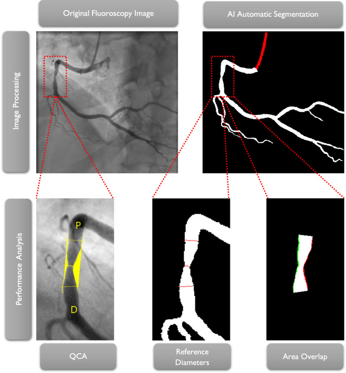
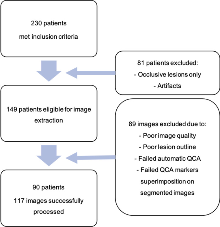
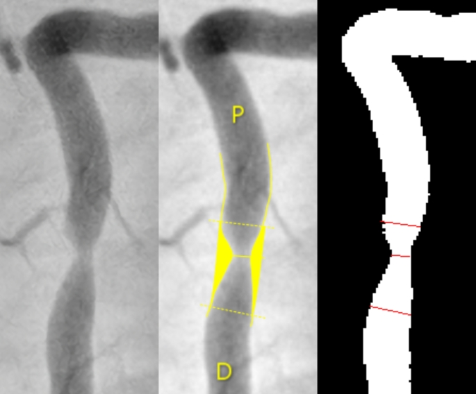
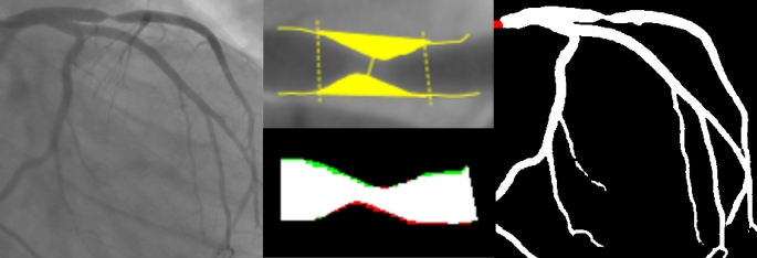
Methods: Retrospective selection of patients undergoing CAG and percutaneous coronary intervention or invasive physiology assessment over a one month period from four centers. A single frame was selected from images containing a lesion with a 50-99% stenosis (visual estimation). Automatic Quantitative Coronary Analysis (QCA) was performed with a validated software. Images were then segmented by the AI model. Lesion diameters, area overlap [based on true positive (TP) and true negative (TN) pixels] and a global segmentation score (GSS - 0 -100 points) - previously developed and published - were measured.
Results: 123 regions of interest from 117 images across 90 patients were included. There were no significant differences between lesion diameter, percentage diameter stenosis and distal border diameter between the original/segmented images. There was a statistically significant albeit minor difference [0,19 mm (0,09-0,28)] regarding proximal border diameter. Overlap accuracy ((TP + TN)/(TP + TN + FP + FN)), sensitivity (TP / (TP + FN)) and Dice Score (2TP / (2TP + FN + FP)) between original/segmented images was 99,9%, 95,1% and 94,8%, respectively. The GSS was 92 (87-96), similar to the previously obtained value in the training dataset.
Conclusion: the AI model was capable of accurate CAG segmentation across multiple performance metrics, when applied to a multicentric validation dataset. This paves the way for future research on its clinical uses.
Keywords: Artificial Intelligence; Coronary angiography; Coronary artery disease; Deep learning; Machine learning; Percutaneous coronary intervention..
© 2023. The Author(s).
Conflict of interest statement
Not applicable.
Figures

Similar articles
-
Development of deep learning segmentation models for coronary X-ray angiography: Quality assessment by a new global segmentation score and comparison with human performance.Rev Port Cardiol. 2022 Dec;41(12):1011-1021. doi: 10.1016/j.repc.2022.04.001. Epub 2022 May 26. Rev Port Cardiol. 2022. PMID: 36511271 English, Portuguese.
-
Automatic coronary artery segmentation and diagnosis of stenosis by deep learning based on computed tomographic coronary angiography.Eur Radiol. 2022 Sep;32(9):6037-6045. doi: 10.1007/s00330-022-08761-z. Epub 2022 Apr 8. Eur Radiol. 2022. PMID: 35394183
-
Non-invasive derivation of instantaneous free-wave ratio from invasive coronary angiography using a new deep learning artificial intelligence model and comparison with human operators' performance.Int J Cardiovasc Imaging. 2025 Apr;41(4):755-771. doi: 10.1007/s10554-025-03369-y. Epub 2025 Mar 10. Int J Cardiovasc Imaging. 2025. PMID: 40063156 Free PMC article.
-
Stenosis Detection and Quantification of Coronary Artery Using Machine Learning and Deep Learning.Angiology. 2024 May;75(5):405-416. doi: 10.1177/00033197231187063. Epub 2023 Jul 3. Angiology. 2024. PMID: 37399509 Review.
-
Current State and Future Perspectives of Artificial Intelligence for Automated Coronary Angiography Imaging Analysis in Patients with Ischemic Heart Disease.Curr Cardiol Rep. 2022 Apr;24(4):365-376. doi: 10.1007/s11886-022-01655-y. Epub 2022 Mar 28. Curr Cardiol Rep. 2022. PMID: 35347566 Free PMC article. Review.
Cited by
-
Old Habits Die Hard: Can AI Help Bring Coronary Angiography Into the 21st Century?JACC Adv. 2024 Jul 4;3(8):101093. doi: 10.1016/j.jacadv.2024.101093. eCollection 2024 Aug. JACC Adv. 2024. PMID: 39055273 Free PMC article.
-
Validation of artificial intelligence-based quantitative coronary angiography.Digit Health. 2024 Dec 18;10:20552076241306937. doi: 10.1177/20552076241306937. eCollection 2024 Jan-Dec. Digit Health. 2024. PMID: 39698508 Free PMC article.
-
Enhancing quantitative coronary angiography (QCA) with advanced artificial intelligence: comparison with manual QCA and visual estimation.Int J Cardiovasc Imaging. 2025 Mar;41(3):559-568. doi: 10.1007/s10554-025-03342-9. Epub 2025 Jan 29. Int J Cardiovasc Imaging. 2025. PMID: 39875702 Free PMC article.
-
Optimizing ensemble U-Net architectures for robust coronary vessel segmentation in angiographic images.Sci Rep. 2024 Mar 19;14(1):6640. doi: 10.1038/s41598-024-57198-5. Sci Rep. 2024. PMID: 38503839 Free PMC article.
-
AI in interventional cardiology: Innovations and challenges.Heliyon. 2024 Aug 26;10(17):e36691. doi: 10.1016/j.heliyon.2024.e36691. eCollection 2024 Sep 15. Heliyon. 2024. PMID: 39281582 Free PMC article. Review.
References
MeSH terms
LinkOut - more resources
Full Text Sources
Miscellaneous
