. 2023 Apr;36(2):279-293.
doi: 10.1007/s10334-023-01077-z. Epub 2023 Apr 7.

High-resolution imaging of the excised porcine heart at a whole-body 7 T MRI system using an 8Tx/16Rx pTx coil

Affiliations

High-resolution imaging of the excised porcine heart at a whole-body 7 T MRI system using an 8Tx/16Rx pTx coil

Maxim Terekhov et al. MAGMA. 2023 Apr.

Abstract

Introduction: MRI of excised hearts at ultra-high field strengths ([Formula: see text]≥7 T) can provide high-resolution, high-fidelity ground truth data for biomedical studies, imaging science, and artificial intelligence. In this study, we demonstrate the capabilities of a custom-built, multiple-element transceiver array customized for high-resolution imaging of excised hearts.

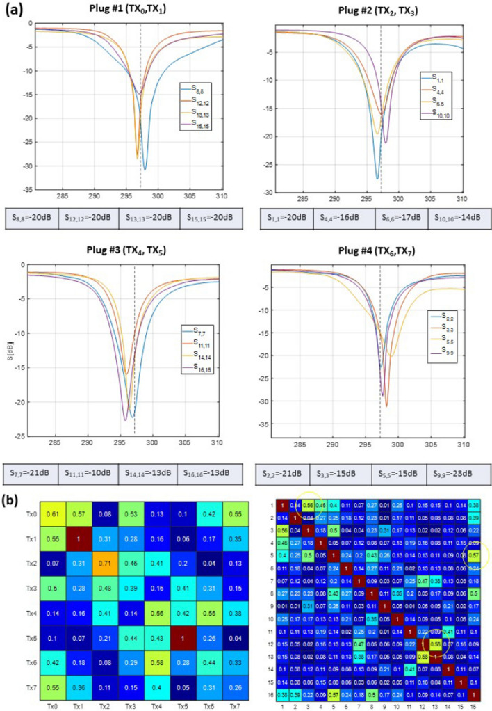
Method: A dedicated 16-element transceiver loop array was implemented for operation in parallel transmit (pTx) mode (8Tx/16Rx) of a clinical whole-body 7 T MRI system. The initial adjustment of the array was performed using full-wave 3D-electromagnetic simulation with subsequent final fine-tuning on the bench.

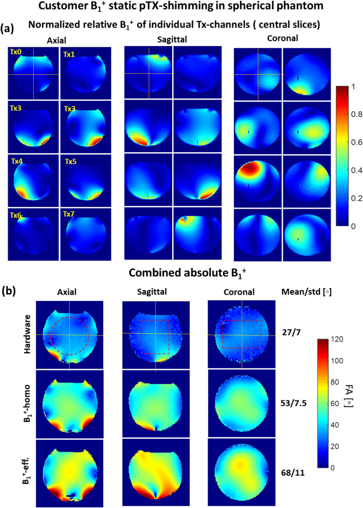
Results: We report the results of testing the implemented array in tissue-mimicking liquid phantoms and excised porcine hearts. The array demonstrated high efficiency of parallel transmits characteristics enabling efficient pTX-based B1+-shimming.

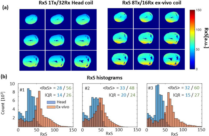
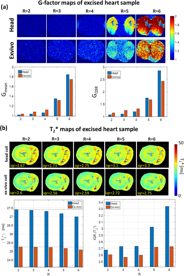
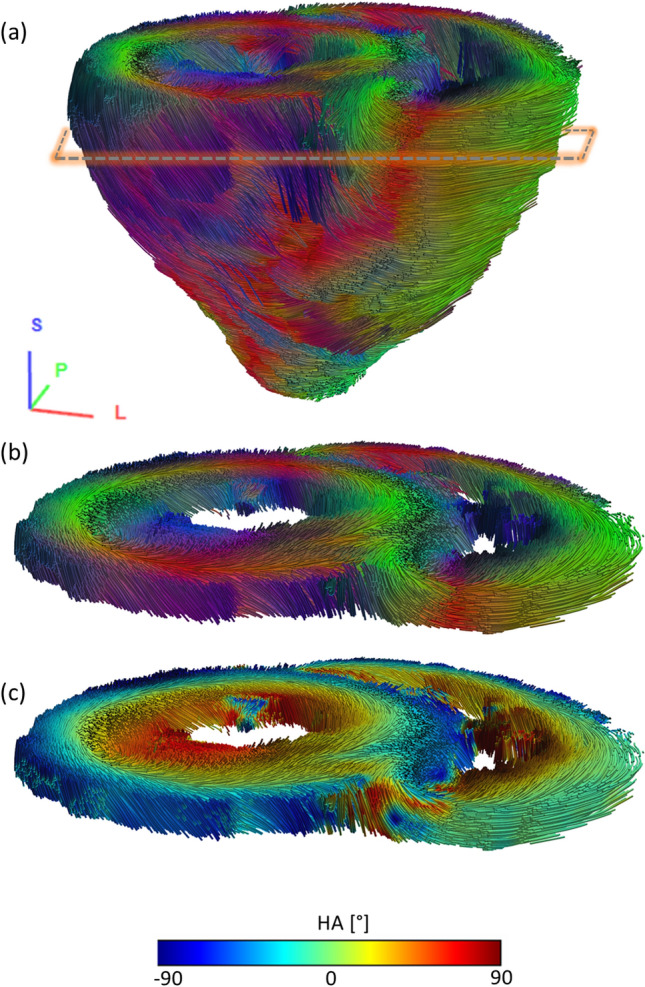
Conclusion: The receive sensitivity and parallel imaging capability of the dedicated coil were superior to that of a commercial 1Tx/32Rx head coil in both SNR and T2*-mapping. The array was successfully tested to acquire ultra-high-resolution (0.1 × 0.1 × 0.8 mm voxel) images of post-infarction scar tissue. High-resolution (isotropic 1.6 mm3 voxel) diffusion tensor imaging-based tractography provided high-resolution information about normal myocardial fiber orientation.

Keywords: Excised heart; Parallel transmit; RF-array; Ultra-high-field.


Conflict of interest statement

The authors declare that they have no conflicts of interest.

Figures

Fig. 1
Fig. 1
Design and schematic of the ex-vivo array for high-resolution imaging of excised porcine hearts. a Coil elements layout, dimensions, and element numbering. b RF simulation model of the array loaded with a 10-cm spherical phantom in front, top, and side views. c A prototype of the 16-element antisymmetric array in the top, bottom, and side views. Phase shifters (PS) and cable traps (CT) are labeled accordingly. Every two neighboring loops are paired to be interfaced to the corresponding Tx channel of RFPA (channel numbering is according to the MRI system notation) to form an 8Tx/16Rx array configuration
Fig. 2
Fig. 2
a Measured S-parameters versus frequency for the four ODU plugs when the array was loaded with a 10-cm spherical phantom (εr= 59.3 and σ=0.79 S/m). Most of the elements were matched better than − 14 dB. Only elements 11 and 16 were matched to − 10 and − 13 dB. b The noise correlation coefficient matrix for 16 receive elements and normalized scattering matrix for 8 Tx channels measured by the MR scanner. Yellow circles on the noise correlation matrix show pairs with relatively high transmission coefficients (> -9 dB) leading to increased noise correlation
Fig. 3
Fig. 3
Static pTX-based B1 + shimming in the homogeneous spherical phantom based on the customer optimization procedure (Appendix 1). (a) Relative normalized B1 + maps of the individual channels used for B1 + optimization. Central slice projections (solid lines) are shown. (b) Combined absolute B1 + maps acquired with default array phasing ("Hardware") and using pTX-based RF-shimming settings computed within the labeled ROI (dashed red lines) by optimization of “B1 + homogeneity” and “B1 + efficiency” cost functions (Appendix 1). All three combined B1 + maps are acquired with the same transmitter reference voltage of 100 V. The mean and standard deviation of the flip angle within shimming ROI are shown for each shimming setting
Fig. 4
Fig. 4
Customer B1 + shimming for the excised heart sample. (a) Normalized relative B1 + -maps of the eight individual transmit channels (one axial slice shown). (b) Normalized combined relative B1-maps before pTx optimization (with hardware phases) and after pTx-based RF-shimming
Fig. 5
Fig. 5
Rx-sensitivity comparison for head coil and ex-vivo coil. (a) Example of Rx-sensitivity maps (9 slices of 32) acquired using ex-vivo array in the heart sample #3 in comparison to the same slices acquired with 1Tx/32Rx head coil. A higher mean value of normalized SNR is observed visually for the ex-vivo array. b Histograms of Rx-sensitivity for 3 ex-vivo heart samples for the head coil (blue bars) and ex-vivo array (red bars). The advantage of the ex-vivo array in filling factor manifests in essentially higher mean Rx-sensitivity. The larger heterogeneity (characterized by interquartile range) of normalized SNR is, however, a consequence of smaller Tx/Rx ex-vivo array volume compared to the resonator of the head coil
Fig. 6
Fig. 6
Noise amplification in parallel acquisition imaging. (a) Maps of the g-factor and statistical metrics for both coils measured in the excised heart sample #3. The mean and maximal (98-percentile) g-factor values become higher for the head coil for the high acceleration factors R = 5,6. b T2* maps of heart sample #3, reconstructed from mGRE images using increasing GRAPPA acceleration factors. There is a remarkable increase of the noise amplification at (R = 5,6) for the head coil leading to 20% increase T2* IQR values within the marked (dashed line) myocardial region when compared with results obtained with the new excised tissue coil
Fig. 7
Fig. 7
Ultra-high spatial resolution images of an excised heart 60 days after infarction. The top row shows a single slice with a whole-heart long-axis view and zoomed region of the scar (labeled by the yellow rectangle). The bottom row shows zoomed regions of individual slices covering the scar region. Images were acquired with parallel receive acceleration factors R = 2 (top row slice thickness 1 mm, acquisition time ~ 55 min) and R = 3 (bottom row, slice thickness 0.8 mm, acquisition time ~ 40 min), respectively. In both cases, a very high level of detail is observed in post-infarction scar tissue
Fig. 8
Fig. 8
Tractography of myofiber bundles based on high-resolution DTI imaging of a fixed porcine heart acquired using the new coil. a Tractography showing 30,000 fiber bundle tracts visualized using tubes with a thickness of 5% of the voxel size. Thresholds for tracking were FA: 0.1, min bundle lengths: 10 mm, max bundle lengths: 300 mm, step size: 0.5 of the voxel size, and angle: 60°. Color-coding of the tracts corresponds to the main eigenvector orientation. A respective coordinate system is given in the bottom left. The dashed box approximates the position of the slab shown in (b) and (c). (b) Excerpt (thickness corresponds to three slices) from the whole-heart tractography. The high resolution enables the visualization and assessment of papillary muscle (dark blue) within the left and right ventricle or the transmural helical configuration of myocyte bundles as well as the intersection of the left and right ventricle with a fidelity unobtainable in-vivo. Color-coding of the tracts corresponds to the main eigenvector orientation as in (a). (c) The same tractography excerpt with color-coding of the tracts corresponds to the local helix angle value

Similar articles

Cited by

  • Ultra-high field cardiac MRI in large animals and humans for translational cardiovascular research.
    Schreiber LM, Lohr D, Baltes S, Vogel U, Elabyad IA, Bille M, Reiter T, Kosmala A, Gassenmaier T, Stefanescu MR, Kollmann A, Aures J, Schnitter F, Pali M, Ueda Y, Williams T, Christa M, Hofmann U, Bauer W, Gerull B, Zernecke A, Ergün S, Terekhov M. Schreiber LM, et al. Front Cardiovasc Med. 2023 May 15;10:1068390. doi: 10.3389/fcvm.2023.1068390. eCollection 2023. Front Cardiovasc Med. 2023. PMID: 37255709 Free PMC article.

References

    1. Van de Moortele PF, Akgun C, Adriany G, Moeller S, Ritter J, Collins CM, Smith MB, Vaughan JT, Ugurbil K. B(1) destructive interferences and spatial phase patterns at 7 T with a head transceiver array coil. Magn Reson Med. 2005;54(6):1503–1518. doi: 10.1002/mrm.20708. - DOI - PubMed
    1. Ibrahim TS, Lee R, Abduljalil AM, Baertlein BA, Robitaille PM. Dielectric resonances and B(1) field inhomogeneity in UHFMRI: computational analysis and experimental findings. Magn Reson Imaging. 2001;19(2):219–226. doi: 10.1016/S0730-725X(01)00300-9. - DOI - PubMed
    1. Xin SX, Huang QH, Gao Y, Li BG, Xu YK, Chen WF. Fetus MRI at 7 T: B-1 shimming strategy and SAR safety implications. IEEE Trans Microw Theory Tech. 2013;61(5):2146–2152. doi: 10.1109/TMTT.2013.2247053. - DOI
    1. Ibrahim TS. Ultrahigh-Field MRI whole-slice and localized RF field excitations using the same RF transmit array. IEEE Trans Med Imaging. 2006;25(10):1341–1347. doi: 10.1109/TMI.2006.880666. - DOI - PubMed
    1. Mao W, Smith MB, Collins CM. Exploring the limits of RF shimming for high-field MRI of the human head. Magn Reson Med. 2006;56(4):918–922. doi: 10.1002/mrm.21013. - DOI - PMC - PubMed

LinkOut - more resources