. 2023 Sep 1;164(9):1965-1975.
doi: 10.1097/j.pain.0000000000002892. Epub 2023 Apr 7.

Local analgesia of electroacupuncture is mediated by the recruitment of neutrophils and released β-endorphins

Affiliations

Local analgesia of electroacupuncture is mediated by the recruitment of neutrophils and released β-endorphins

Jing-Tao Shi et al. Pain. .

Abstract

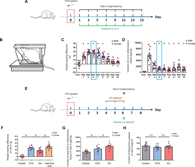
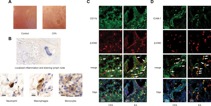
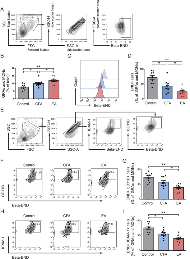
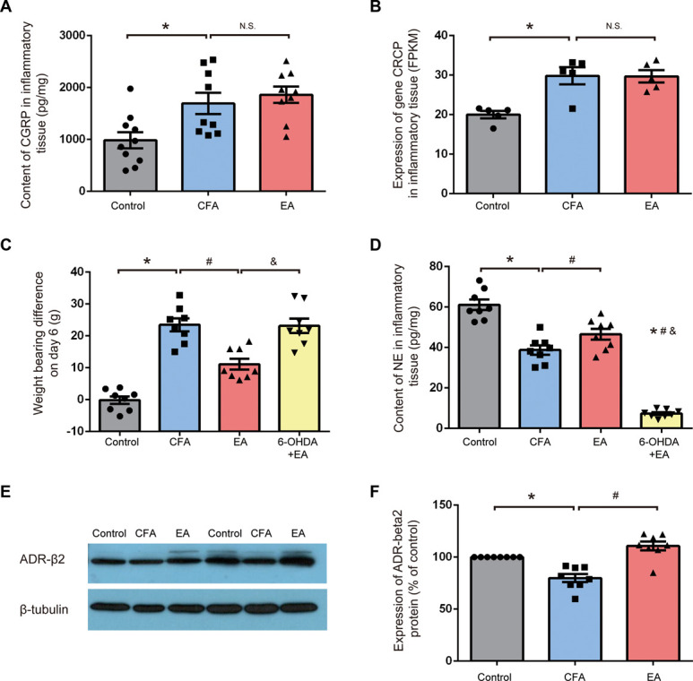
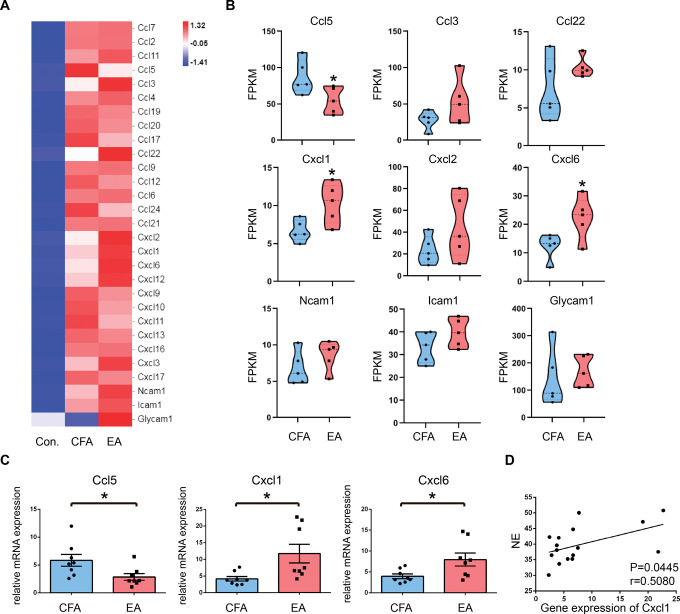
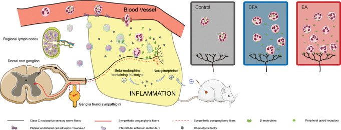
The efficacy of acupuncture in treating pain diseases has been recognized in clinical practice, and its mechanism of action has been a hot topic in academic acupuncture research. Previous basic research on acupuncture analgesia has focused mostly on the nervous system, with few studies addressing the immune system as a potential pathway of acupuncture analgesia. In this study, we investigated the effect of electroacupuncture (EA) on the β-endorphins (β-END) content, END-containing leukocyte type and number, sympathetic neurotransmitter norepinephrine (NE), and chemokine gene expression in inflamed tissues. To induce inflammatory pain, about 200 µL of complete Frester adjuvant (CFA) was injected into the unilateral medial femoral muscle of adult Wistar rats. Electroacupuncture treatment was performed for 3 days beginning on day 4 after CFA injection, with parameters of 2/100 Hz, 2 mA, and 30 minutes per treatment. The weight-bearing experiment and enzyme-linked immunosorbent assay showed that EA treatment significantly relieved spontaneous pain-like behaviors and increased the level of β-END in inflamed tissue. Injection of anti-END antibody in inflamed tissue blocked this analgesic effect. Flow cytometry and immunofluorescence staining revealed that the EA-induced increase in β-END was derived from opioid-containing ICAM-1 + /CD11b + immune cells in inflamed tissue. In addition, EA treatment increased the NE content and expression of β2 adrenergic receptor (ADR-β2) in inflammatory tissues and upregulated Cxcl1 and Cxcl6 gene expression levels. These findings provide new evidence for the peripheral analgesic effect of acupuncture treatment by recruiting β-END-containing ICAM-1 + /CD11b + immune cells and increasing the β-END content at the site of inflammation.


Conflict of interest statement

The authors have no conflict of interest to declare.

Sponsorships or competing interests that may be relevant to content are disclosed at the end of this article.

Figures

Figure 1.
Figure 1.
EA treatment increased the content of β-END in inflammatory tissues and relieved pain. (A) Experimental procedure timeline of observation of pain and local inflammatory reaction after intramuscular injection of CFA. N = 6 male and 6 female Wistar rats per group, total of 9 groups: control, day 1, day 3, day5, day 7, day 9, day 11, day 13, and day15. Injections were given intramuscularly. (B) A schematic illustration of behavioral testing. Changes in weight-bearing difference (C) and Interleukin- 6 (IL-6) levels in inflammatory tissues (D) measured at 15 days after CFA injection, *P < 0.05 vs control group, #P < 0.05 vs day 1. (E) Experimental procedure timeline of EA treatment. N = 6 male and 6 female Wistar rats per group, total of 4 groups: control, CFA, EA, and Anti-End + EA (antibody concentration: 8 mg/mL, 110 µL). (F) Weight-bearing difference was tested on day 6 after CFA injection in the indicated groups, *P < 0.05 vs control group, #P < 0.05 vs CFA group, &P < 0.05 vs EA group. β-END levels in the tissues (G) and plasma (H) were measured on day 6 after CFA injection and EA treatment, *P < 0.05 vs control group, #P < 0.05 vs CFA group. All data are shown as mean ± SEM; statistical analyses were performed using one-way ANOVA followed by the Tukey honestly significant difference (Tukey HSD) test. ANOVA, analysis of variance; β-END, β-endorphins; CFA, complete Frester adjuvant; EA, electroacupuncture.
Figure 2.
Figure 2.
EA treatment increased the number of β-END containing ICAM-1+/CD11b+ immune cells in inflammatory tissue. (A) Representative examples of muscles from rats after CFA injection and control treatment. (B) Immunohistochemistry images of sections stained with β-END showing inflammatory cell infiltration. Dark brown staining represents β-END immunoreactivity. Neutrophils exhibited a typical polymorphic nucleus, macrophages exhibited an elongated or indented oval nucleus, and monocytes exhibited an oval notched or horseshoe-shaped nucleus. (C and D) Immunofluorescence staining of inflammatory tissue using a mouse anti-β-END antibody (red), rabbit anti-ICAM-1 antibody or anti-CD11b antibody (all were marked green), and DAPI. Arrows point at double-positive cells. β-END, β-endorphins; CFA, complete Frester adjuvant; EA, electroacupuncture.
Figure 3.
Figure 3.
EA treatment increased proportions of GRAs and MONs and decreased the proportions of β-END+ CD11b+ and β-END+ ICAM-1+ cells on day 6 after CFA injection. (A) Flow cytometry plots showed the gating strategy for β-END+ cells in GRAs and MONs. (B) Bar graph showed the change of GRAs and MONs (% of total) on day 6 after CFA injection in 3 groups (n = 9). (C and D) Flow cytometry plots and bar graph show the expression of β-END in GRAs and MONs in 3 groups (n = 9). (E) Gating strategy of β-END+ ICAM-1+ cells and β-END+ CD11b+ cells in GRAs and MONs. Flow cytometry plots and bar graphs show the proportions of β-END+ CD11b+ (F and G) and β-END+ ICAM-1+ (H and I) cells in GRAs and MONs in 3 groups. **P < 0.01 vs control group, #P < 0.05 vs CFA group. All data are shown as mean ± SEM; statistical analyses were performed using 1-way ANOVA followed by the Tukey HSD test. ANOVA, analysis of variance; β-END, β-endorphins; CFA, complete Frester adjuvant; EA, electroacupuncture; FSC, forward scatter; FSC-A, forward scatter area; GRAs, granulocytes; MONs, monocytes; SSC, side scatter; SSC-A, side scatter area; SSC-H, side scatter height.
Figure 4.
Figure 4.
Sympathetic nerve mediating the analgesic effect of EA treatment. (A) Compared with the control group, content of CGRP in inflammatory tissue was higher in the CFA group, but it was not significantly different between the CFA group and EA group (n = 9). (B) Expression of gene Crcp in FPKM was generated from whole-transcriptome sequencing in result 5. Compared with the control group, expression of gene Crcp in inflammatory tissue was higher in the CFA group, but it was not significantly different between the CFA group and EA group (n = 5). (C) Contents of NE was measured on day 6 after CFA injection in the control, CFA, EA, and 6-OHDA + EA groups (n = 8). *P < 0.05 vs control group, #P < 0.05 vs CFA group, &P < 0.05 vs EA group. (D) Weight-bearing difference was tested on day 6 after CFA injection in the control, CFA, EA, and 6-OHDA + EA groups (n = 8). *P < 0.05 vs control group, #P < 0.05 vs CFA group, &P < 0.05 vs EA group. (E) Expression of β2 adrenergic receptor as determined by the Western blotting test. (F) Compared with the control group, ADR-β2 expression in inflammatory tissue was lower in the CFA group. ADR-β2 expression in the EA group was significantly higher compared with the CFA group (n = 8). *P < 0.05 vs control group, #P < 0.05 vs CFA group. All data are shown as mean ± SEM; statistical analyses were performed using 1-way ANOVA followed by the Tukey HSD test. ADR-β2, β2 adrenergic receptor; ANOVA, analysis of variance; CFA, complete Frester adjuvant; CGRP, calcitonin gene-related peptide; EA, electroacupuncture; FPKM, fragments per kb of transcript per million fragments mapped; NE, norepinephrine; OHDA, hydroxydopamine.
Figure 5.
Figure 5.
RNA-sequencing analysis and qRT-PCR verification of genes related to cell migration. (A) Heat map of cell migration–associated genes. Different heat map colors represent the relative mRNA expression levels of genes calculated by Log2 (FPKM + 1). (B) Violin plots of expression values for differentially expressed genes between CFA and EA groups. Results are expressed as FPKM. Data shown are the mean FPKM  ±  SEM (n = 5). *P < 0.05 vs CFA group. (C) Significant differentially expressed genes as determined by real-time PCR (n = 8). (D) Correlation between NE and the gene expression of Cxcl1 in inflammatory tissues based on the Pearson correlation analysis. All data are expressed as the mean ± SEM. *P < 0.05 vs CFA group. CFA, complete Frester adjuvant; EA, electroacupuncture; FPKM, fragments per kb of transcript per million fragments mapped; NE, norepinephrine.
Figure 6.
Figure 6.
EA exerts a local analgesic effect during peripheral inflammation by activating sympathetic nerve fibers and promoting the migration of β-END containing immune cells to the pain site. β-END, β-endorphins; CFA, complete Frester adjuvant; EA, electroacupuncture.

References

    1. Akerman S, Kaube H, Goadsby PJ. Vanilloid type 1 receptors (VR1) on trigeminal sensory nerve fibres play a minor role in neurogenic dural vasodilatation, and are involved in capsaicin-induced dural dilation. Br J Pharmacol 2003;140:718–24. - PMC - PubMed
    1. Binder W, Mousa SA, Sitte N, Kaiser M, Stein C, Schäfer M. Sympathetic activation triggers endogenous opioid release and analgesia within peripheral inflamed tissue. Eur J Neurosci 2004;20:92–100. - PubMed
    1. Bing Z, Villanueva L, Le Bars D. Acupuncture and diffuse noxious inhibitory controls: naloxone-reversible depression of activities of trigeminal convergent neurons. Neuroscience 1990;37:809–18. - PubMed
    1. Brack A, Labuz D, Schiltz A, Rittner HL, Machelska H, Schafer M, Reszka R, Stein C. Tissue monocytes/macrophages in inflammation: hyperalgesia versus opioid-mediated peripheral antinociception. Anesthesiology 2004;101:204–11. - PubMed
    1. Cabot PJ, Carter L, Schafer M, Stein C. Methionine-enkephalin-and Dynorphin A-release from immune cells and control of inflammatory pain. PAIN 2001;93:207–12. - PubMed

Publication types