. 2023 Apr 11;120(15):e2201910120.
doi: 10.1073/pnas.2201910120. Epub 2023 Apr 7.

High-resolution structural information of membrane-bound α-synuclein provides insight into the MoA of the anti-Parkinson drug UCB0599

Affiliations

High-resolution structural information of membrane-bound α-synuclein provides insight into the MoA of the anti-Parkinson drug UCB0599

Thomas C Schwarz et al. Proc Natl Acad Sci U S A. .

Abstract

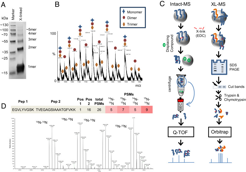
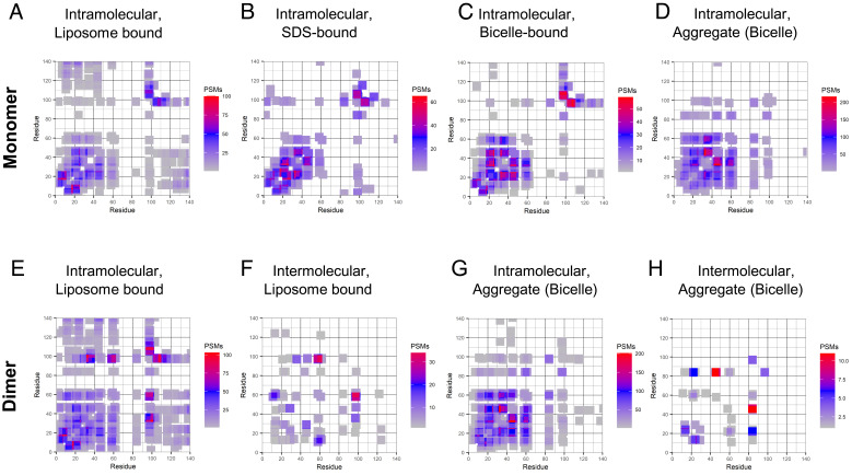
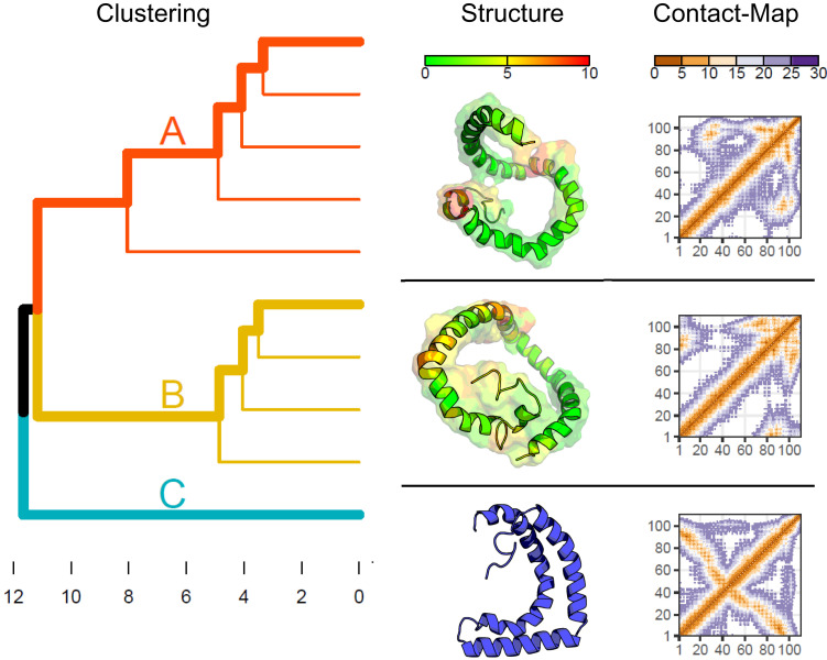
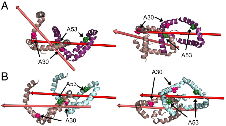
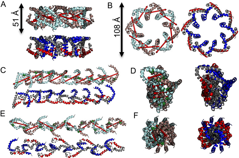
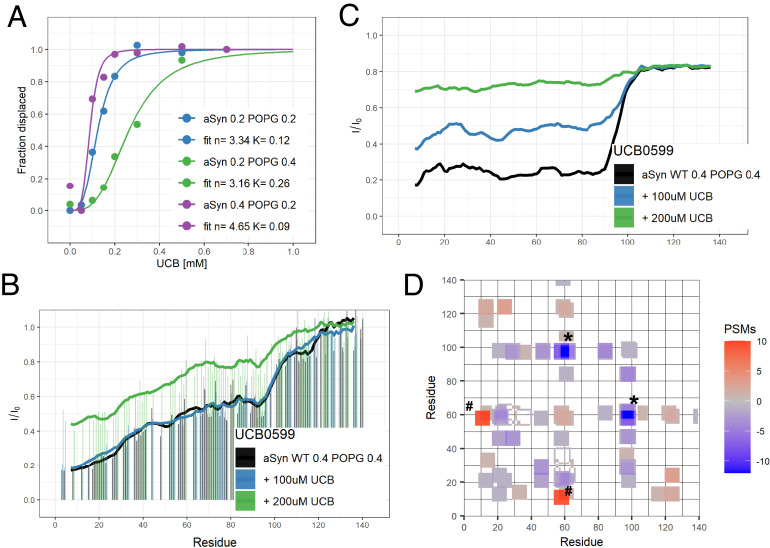
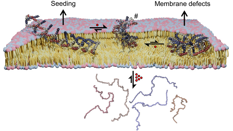
α-synuclein (αS) is an intrinsically disordered protein whose functional ambivalence and protein structural plasticity are iconic. Coordinated protein recruitment ensures proper vesicle dynamics at the synaptic cleft, while deregulated oligomerization on cellular membranes contributes to cell damage and Parkinson's disease (PD). Despite the protein's pathophysiological relevance, structural knowledge is limited. Here, we employ NMR spectroscopy and chemical cross-link mass spectrometry on 14N/15N-labeled αS mixtures to provide for the first time high-resolution structural information of the membrane-bound oligomeric state of αS and demonstrate that in this state, αS samples a surprisingly small conformational space. Interestingly, the study locates familial Parkinson's disease mutants at the interface between individual αS monomers and reveals different oligomerization processes depending on whether oligomerization occurs on the same membrane surface (cis) or between αS initially attached to different membrane particles (trans). The explanatory power of the obtained high-resolution structural model is used to help determine the mode-of-actionof UCB0599. Here, it is shown that the ligand changes the ensemble of membrane-bound structures, which helps to explain the success this compound, currently being tested in Parkinson's disease patients in a phase 2 trial, has had in animal models of PD.

Keywords: UCB0599; XL-MS; oligomeric structure; paramagnetic NMR spectroscopy; α-synuclein.


Conflict of interest statement

T.S.B. and R.J.T. were employed by UCB Pharma and held stock options and shares of the same during the writing of this paper. T.C.S. and R.K. received funding from UCB Pharma and Neuropore Therapies. T.C.S., K.L., and R.K. are co-authors on a publication describing an early molecule showing a “proof-of mechanism” used by UCB0599.

Figures

Fig. 1.
Fig. 1.
Cross-linking of α-synuclein at the membrane. (A) SDS-PAGE (4 to 20%) of αS cross-linked in the presence of POPG-based liposomes and subsequent removal of liposomes followed by SEC. Molecular mass increases from monomers up to tetramers are clearly visible. Higher oligomers are present to a smaller degree. (B) Selection of m/z species showing the masses of monomeric, dimeric, and trimeric forms of cross-linked αS as detected by intact mass spectrometry post SEC. (C) Schemes for cross-linking and purification as used for detection of oligomers in intact mass (Left) and intermolecular/intramolecular cross-links of peptides (Right). A previously reported molecule that releases αS from membrane surfaces (37) is depicted as a green hexagon. (D) Spectrum demonstrating how inter- and intra-molecular cross-links can be distinguished through isotopomeric labeling. At the Top, the two different peptides detected are indicated with their cross-linking positions as well as the detected peptide spectrum matches for each combination of isotopomers. The mass spectrum allowing for this distinction is shown below.
Fig. 2.
Fig. 2.
Cross-linking patterns in α-synuclein are sensitive to conditions used. (AD) Cross-links detected in the monomer-band of αS after incubation with various membrane mimics. The number of PSMs found in a four-residue window is indicated in the color scales. While some long-distance contacts (130–20) are observed in the liposome-bound monomer (A) and medium distances (60–35) in bicelles (C), SDS bound monomers do not show a significant amount of long-range interactions (B). Both intra- and inter-molecular contacts were detected for the bound form from the dimer band of αS (E and F). Interestingly, the intramolecular cross-linking pattern changed substantially between dimeric, bound αS (E) and the monomeric state (A) (statistical measures are available in SI Appendix, Fig. S22). Intermolecular contacts detected indicate two main regions for dimer formation (60–15 and 95–60). Note that loop-links (cross-links within a single peptide fragment) were omitted in our analysis, thus no diagonal peaks are expected. Additionally, residues located in the segment 60–80 are not amenable to the cross-linking reagents used and therefore no cross-links are observed for these residues. (G and H) When incubated with bicelles αS can form aggregates. These show dissimilar cross-linking patterns as compared to those on liposomes. Intramolecular contacts both in the monomer as well as the dimer bands (D and G) were more plentiful and show fewer medium range contacts (95–60 and 95–35). Intermolecular contacts (H), however, are less abundant and are changed drastically as compared to the liposome-bound dimer.
Fig. 3.
Fig. 3.
The PRE/PRI/SSP derived compact ensemble of α-synuclein shares features with the multimer forming ensemble at the membrane. (Left) CA-CA rmsd clustering of the 10 lowest energy structures obtained from monomer calculations. Residues 101 to 140 are excluded from the analysis as they are not constrained by experimental data. Three major clusters are indicated by different colors (A: red, B: dark yellow and C: light blue), the cluster representatives are highlighted by bold lines, rmsd [Å] is indicated at the bottom. (Middle) Representative 3D cluster structures. The surface representation includes all structures contained in the cluster aligned to the cluster representative and is colored by the maximum CA-CA distance between structures at each position. Since cluster C contains only a single structure, no common surface representation is given. (Right) CA-CA based contact map for the individual clusters. The color codes for the maximum distance in surface representations (Middle) and the distances in contact maps (Right) are indicated at the Top of each column (both given in [Å]).
Fig. 4.
Fig. 4.
Dimer structures derived from intermolecular XL-MS data. (A and B) Two prototypical solutions of the dimer calculation resembling monomer cluster A. While the interface is similar in both cases, slight deviations lead to a noticeable kink between subunits as demonstrated by the major axis of the monomers. While (A) shows an angle between the major axis of the two subunits (B) shows a near parallel offset. Mutations associated with familial forms of Parkinson’s disease and studied by XL-MS here are indicated by pink (A30) and green (A53) spheres and are located at the interface.
Fig. 5.
Fig. 5.
Propagation of α-synuclein into higher oligomers. Deviations of the interface demonstrated in Fig. 4 lead to distinct oligomeric structures upon further addition of monomers. Dependent on the kink between the major axes, this results in either circular (A and B) or linear/helical/extended (CF) oligomeric structures. Individual αS molecules in the oligomers are coloured alternatingly (cyan brown) in one representation for each view [Top (A, C, and E) and Left (B, D, and F)]. Regions identified to interact with the polar head groups of the membrane surface (1–36: blue) or lipid tails within the inner layer of the lipid bilayer (70–88: red) in ref.  are indicated (Bottom (A, C, and E) and Right (B, D, and F)]. Ring-like structures have a height and diameter of 51 Å and 108 Å, respectively, which is in good agreement with dimensions extracted from EM-particle classifications. (CF) Two representative structures of extended oligomers. While both oligomers form extended chains upon continuous addition of monomers, the orientation of monomers is different. Small (C and D) or large (E and F) rotational angles result in distinctly different interfaces either forming an amphiphilic-like [surface (1–36, blue) and inner layer (70–88, red) interactions on opposite sides (C and D)] or alternating [surface and inner layer interactions are alternating (E and F)] membrane interaction interface.
Fig. 6.
Fig. 6.
The effect of UCB0599 on α-synuclein. Addition of the small-molecule UCB0599 to liposome-bound αS modifies the underlying ensemble at low concentration and displaces the protein at elevated ligand concentration. (A) Following the stepwise addition of the compound while referencing to the free C-terminus of the protein (SI Appendix, Fig. S5), concentration-dependent displacement of αS from POPG-based liposomes is observed. Here, signals of unbound αS regions are measured and their relative intensity between conditions with and without liposomes reports on the bound fraction. This dependence shows cooperative behavior and is dependent on the ratio between protein and liposomes. Best-HSQC (B) and DEST (C) measurements show that at lower concentrations no change in displaced αS is observed in HSQCs while an increase in DEST intensity already reflects an increase in flexibility of the bound form. When testing for changes in the bound form of the protein (D), clear changes in the cross-linking pattern are observed upon addition of the compound. Intermolecular PSMs show a reduction in links in some regions [(92–101) to (58–62), marked *] while increasing in others [(54–61) to (8–16), marked #]. These regions are found at the interface in our calculated dimer (Fig. 4A) and highlighted in SI Appendix, Fig. S7. The number of PSMs found in a four-residue window is indicated in the color scale.
Fig. 7.
Fig. 7.
Model for membrane-bound oligomeric α-synuclein and UCB0599’s mode-of-action. The membrane-bound state of αS is characterized by an ensemble of different oligomer topologies (circular vs. elongated). While the elongated form (Left) might be involved in seeding with membranes (7) when growing too large, the circular structure (Right) is likely relevant for membrane defects and possibly pore formation (43). Proper functioning of αS requires a subtle balancing in order to avoid the formation of these toxic variants. The proposed MoA of UCB0599 involves interference with oligomeric αS on the membrane and thereby shifting the equilibrium away from species capable of generating toxic effects (elongated and circular) toward a conformational state (represented by the central cartoon marked #) characterized by increased flexibility and decreased membrane embedding. The resulting loosening of membrane-attachment facilitates displacement of αS from the membrane with UCB0599 (depicted as red spheres). At sufficiently high concentrations, αS is displaced from the membrane in a monomeric form (Bottom). Regions of αS interacting with lipid tails (red) or hydrophilic head groups (blue) are indicated (32).

Similar articles

Cited by

References

    1. Goedert M., Jakes R., Spillantini M. G., The synucleinopathies: Twenty years on. J. Parkinsons Dis. 7, S51–S69 (2017). - PMC - PubMed
    1. Alam P., Bousset L., Melki R., Otzen D. E., α-synuclein oligomers and fibrils: A spectrum of species, a spectrum of toxicities. J. Neurochem. 150, 522–534 (2019). - PubMed
    1. Wong Y. C., Krainc D., alpha-synuclein toxicity in neurodegeneration: Mechanism and therapeutic strategies. Nat. Med. 23, 1–13 (2017). - PMC - PubMed
    1. van Maarschalkerweerd A., Vetri V., Langkilde A. E., Fodera V., Vestergaard B., Protein/lipid coaggregates are formed during alpha-synuclein-induced disruption of lipid bilayers. Biomacromolecules 15, 3643–3654 (2014). - PubMed
    1. Weinreb P. H., Zhen W., Poon A. W., Conway K. A., Lansbury P. T. Jr., NACP, a protein implicated in Alzheimer’s disease and learning, is natively unfolded. Biochemistry 35, 13709–13715 (1996). - PubMed

Publication types

MeSH terms