. 2023 Jul 1;139(1):49-62.
doi: 10.1097/ALN.0000000000004579.

Measures of Information Content during Anesthesia and Emergence in the Caenorhabditis elegans Nervous System

Affiliations

Measures of Information Content during Anesthesia and Emergence in the Caenorhabditis elegans Nervous System

Andrew S Chang et al. Anesthesiology. .

Abstract

Background: Suppression of behavioral and physical responses defines the anesthetized state. This is accompanied, in humans, by characteristic changes in electroencephalogram patterns. However, these measures reveal little about the neuron or circuit-level physiologic action of anesthetics nor how information is trafficked between neurons. This study assessed whether entropy-based metrics can differentiate between the awake and anesthetized state in Caenorhabditis elegans and characterize emergence from anesthesia at the level of interneuronal communication.

Methods: Volumetric fluorescence imaging measured neuronal activity across a large portion of the C. elegans nervous system at cellular resolution during distinct states of isoflurane anesthesia, as well as during emergence from the anesthetized state. Using a generalized model of interneuronal communication, new entropy metrics were empirically derived that can distinguish the awake and anesthetized states.

Results: This study derived three new entropy-based metrics that distinguish between stable awake and anesthetized states (isoflurane, n = 10) while possessing plausible physiologic interpretations. State decoupling is elevated in the anesthetized state (0%: 48.8 ± 3.50%; 4%: 66.9 ± 6.08%; 8%: 65.1 ± 5.16%; 0% vs. 4%, P < 0.001; 0% vs. 8%, P < 0.001), while internal predictability (0%: 46.0 ± 2.94%; 4%: 27.7 ± 5.13%; 8%: 30.5 ± 4.56%; 0% vs. 4%, P < 0.001; 0% vs. 8%, P < 0.001), and system consistency (0%: 2.64 ± 1.27%; 4%: 0.97 ± 1.38%; 8%: 1.14 ± 0.47%; 0% vs. 4%, P = 0.006; 0% vs. 8%, P = 0.015) are suppressed. These new metrics also resolve to baseline during gradual emergence of C. elegans from moderate levels of anesthesia to the awake state (n = 8). The results of this study show that early emergence from isoflurane anesthesia in C. elegans is characterized by the rapid resolution of an elevation in high frequency activity (n = 8, P = 0.032). The entropy-based metrics mutual information and transfer entropy, however, did not differentiate well between the awake and anesthetized states.

Conclusions: Novel empirically derived entropy metrics better distinguish the awake and anesthetized states compared to extant metrics and reveal meaningful differences in information transfer characteristics between states.


Conflict of interest statement

Conflicts of Interest

None.

Figures
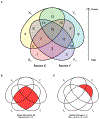
Figure 1
Figure 1
A four-signal model for entropy-based information transfer between neuronal pairs. A) Venn diagram of the four-signal model of past and future information content of two sources X and Y. The 15 regions are numbered by binary combination of XP, YP, XF and YF with values of 1, 2, 4 and 8 respectively. The region colors are assigned according to shading combinations of the base colors shown in regions 1, 2, 4 and 8. B) The entropy quantity of Mutual Information (MI), representing the information that is common between the past states of source X and Y. Graphically, this corresponds to the intersection of the regions XP and YP, i.e. the combination of regions 3, 7, 11, 15, described in set notation as XPYP, whose information content is therefore H(XP)+H(YP)H(XP,YP). C) The entropy quantity of Transfer Entropy from X to Y (TEXY), representing the information that is present in the future state of Y (YF) that can be predicted from the past of X (XP) but not the past of Y (YP), and therefore represents causal transmission of information from X to Y. Graphically, this corresponds to the intersection of regions XP and YF but not YP, i.e. the combination of regions 9 and 13 described in set notation as XP(YFYP), whose information content is therefore H(XP,YP)+H(YP,YF)H(XP,YP,YF)H(YP).
Figure 2
Figure 2
Isoflurane exposure suppresses power and Shannon entropy of neuronal activity signals, and early emergence from isoflurane exposure is characterized by a quickly resolving high-frequency spectral shift. Aabc) Fluorescent optical activity of 120 neurons in the head of C. elegans equilibrated to atmospheres of 0%, 4% and 8% isoflurane respectively. Color represents normalized GCaMP6s fluorescence activity in a cytoplasmic shell surrounding each tracked neuronal nucleus. Ad) Decreasing total signal power under increasing concentrations of isoflurane. Mean signal power ± SD of neuronal activity traces recorded from animals equilibrated to room air, 4% or 8% isoflurane. Ae) Mean Shannon entropy ± SD of quantized neuronal activity traces recorded from animals equilibrated to room air, 4% or 8% isoflurane. Bab) Intensity traces of neuronal activity in 120 neurons in the C. elegans head ganglia before isoflurane exposure and as the animal emerges from equilibration to 4% isoflurane over 120 minutes. Bc) Mean power spectral density of the time-differentiated neuronal activity traces 0.2 and 0.8-hours post-exposure to either room air or 4% isoflurane. Bd) Mean spectral median power frequency (MPF) ± SD of neuronal traces in animals equilibrated to room air or 4% isoflurane, calculated at 12-minute epochs post-exposure. Error bars show the standard error of the mean. (* P < 0.05, ** P < 0.01, *** P < 0.001)
Figure 3
Figure 3
Exposure to isoflurane significantly alters the distribution of entropy within the 4-signal information transfer model. Mean entropy content ± SD in information regions 1-15 of the 4-signal information transfer model in animals exposed to room air, 4%, or 8% isoflurane (n=10). Entropy content was calculated for each neuron pair recorded in each animal (14400 neuron pairs/animal/exposure condition), normalized to the total joint entropy of the 4-signal model, and then averaged by exposure condition. Error bars show the standard error of the mean.
Figure 4
Figure 4
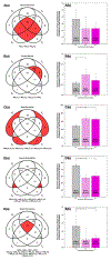
The anesthetized state can be characterized by shifts in novel entropy-based measures of information transfer between neuron pairs. Aa-Ea) Selected information region clusters in the 4-signal model representative of information theoretic metrics: Mutual Information, Transfer Entropy, State Decoupling, Internal Predictability, and System Consistency. Colored Venn areas represent the combined information regions that compose each metric. The equations used to calculate each metric from joint entropies are also shown. Ab-Eb) Mean proportional entropy content ± SD in the selected information region clusters in neuronal activity trace pairs recorded from animals exposed to room air, 4% and 8% isoflurane. Error bars show the standard error of the mean. (* P < 0.05, ** P < 0.01, *** P < 0.001)
Figure 5
Figure 5
Alterations in entropy-based measures of neuronal-pair information transfer resolve to baseline levels non-linearly as animals emerge from isoflurane anesthesia: respectively Mutual Information, Transfer Entropy, State Decoupling, Internal Predictability, and System Consistency. A-E) Time-smoothed means ± 95% CI of proportional entropy content in information region clusters corresponding to measures of information transfer in animals emerging from anesthesia with isoflurane 4% and in room-air exposed controls. Metrics as calculated for individual animals are also shown for each measurement type. (n=8 isoflurane exposed, n=8 controls).

Comment in

References

    1. Shannon CE: A mathematical theory of communication. The Bell System Technical Journal 1948; 27: 379–423
    1. Connor CW: Artificial Intelligence and Machine Learning in Anesthesiology. Anesthesiology 2019; 131: 1346–1359 - PMC - PubMed
    1. Hudetz AG, Pillay S, Wang S, Lee H: Desflurane Anesthesia Alters Cortical Layer–specific Hierarchical Interactions in Rat Cerebral Cortex. Anesthesiology 2020; 132: 1080–1090 - PMC - PubMed
    1. Lipping T, Saareleht J, Olejarczyk E, Sonkajärvi E, Ylinen T, Jäntti V: Synchronization of brain activity during induction of propofol anesthesia: Comparison of methods, 38th Annual International Conference of the IEEE Engineering in Medicine and Biology Society, 2016, pp 5505–5508 - PubMed
    1. Prince LY, Tran MM, Grey D, Saad L, Chasiotis H, Kwag J, Kohl MM, Richards BA: Neocortical inhibitory interneuron subtypes are differentially attuned to synchrony- and rate-coded information. Communications Biology 2021; 4: 935. - PMC - PubMed

Publication types