. 2023 Jun 13;56(6):1239-1254.e7.
doi: 10.1016/j.immuni.2023.03.008. Epub 2023 Apr 6.

Long-term tolerance to skin commensals is established neonatally through a specialized dendritic cell subgroup

Affiliations

Long-term tolerance to skin commensals is established neonatally through a specialized dendritic cell subgroup

Antonin Weckel et al. Immunity. .

Abstract

Early-life establishment of tolerance to commensal bacteria at barrier surfaces carries enduring implications for immune health but remains poorly understood. Here, we showed that tolerance in skin was controlled by microbial interaction with a specialized subset of antigen-presenting cells. More particularly, CD301b+ type 2 conventional dendritic cells (DCs) in neonatal skin were specifically capable of uptake and presentation of commensal antigens for the generation of regulatory T (Treg) cells. CD301b+ DC2 were enriched for phagocytosis and maturation programs, while also expressing tolerogenic markers. In both human and murine skin, these signatures were reinforced by microbial uptake. In contrast to their adult counterparts or other early-life DC subsets, neonatal CD301b+ DC2 highly expressed the retinoic-acid-producing enzyme, RALDH2, the deletion of which limited commensal-specific Treg cell generation. Thus, synergistic interactions between bacteria and a specialized DC subset critically support early-life tolerance at the cutaneous interface.

Keywords: Staphylococcus epidermidis; commensal bacteria; commensals; dendritic cells; early-life immunity; neonatal immunity; regulatory T cells; retinoic acid; skin; tolerance.


Conflict of interest statement

Declaration of interests T.C.S. is on the Scientific Advisory Board of Concerto Biosciences. N.A. is on the Scientific Advisory Board for Shennon Biotech, and consults or lectures for 23andMe, Cellino Biotech, Janssen Pharmaceuticals, and Immunitas Therapeutics.

Figures
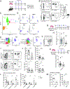
Figure 1:
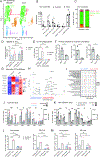
Figure 1:. CD301b+ DC2 are required for early life generation of skin commensal-specific Treg cells
(A) Adult and neonatal mice were colonized with S. epi-2w 3 times every 3 days then harvested a week later; (B) Gating of 2w+CD44+CD4+ T cells (2w+ CD4+ T cells) and 2w+CD44+FoxP3+CD4+ Treg (2w+ Treg cells) (left), and percentage of 2w+ Treg cells in skin draining lymph nodes (SDLN) (pre-gating: live Lin(CD11b, CD11c, F4/80, B220)TCRβ+CD4+). Data from 2–3 independent experiments. (C) UMAP and (D) major defining genes of DC neonatal skin DCs subsets by CITE-seq. (E) Expression of Mgl2 (left) and its protein CD301b (right). (F) Gating for CD301b on D11 skin DC2 (pre-gating: live CD45+CD11c+MHCII+CD103EpCam). (G) Experimental setup for (H-J). (H) Fold-change vs. littermate controls of total 2w+ Treg cells in the indicated mouse models. One sample Wilcoxon test to hypothetical value of 1. Data from 2–3 independent experiments. (I) Gating of 2w+ Treg cells and (J) Percentage and total numbers of 2w+ Treg cells and 2w+CD44+CD4FoxP3CD4+ Teff cells (2w+ Teff cells) in the SDLN of DT-treated Mgl2WT and Mgl2DTR mice at D21. (K) Experimental set-up and quantification of 2w+ Treg cells in SDLN in of Mgl2creH2ab1fl/fl and H2ab1fl/fl littermates. Pooled data from 3 independent experiments. (L) Percentage (left) and total numbers (right) of 2w+ Treg cells in the SDLN of D21 mice neonatally colonized by 2w-expressing coagulase negative staphylococci (CoNS). Pooled data from two independent experiments. Two-way ANOVA. (M) Percentage (left) and total numbers (right) of 2w+ Treg cells in SDLN of D21 mice topically treated with solubilized 2w peptide. One representative experiment of two is shown. Dots represent individual mice. Unless specified Mann-Whitney test was used with *p < 0.05, **p < 0.01, ***p < 0.001, ****p<0.0001, ns = not significant.
Figure 2:
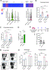
Figure 2:. Neonatal CD301b+ DC2 are required for establishment of long-term tolerance to skin commensals
(A) Mice were treated neonatally with DT and S. epi-2w (as in Fig. 1G), aged out for 2 weeks, then re-colonized concurrent with skin barrier disruption. (B) Total numbers of polyclonal CD4+ (left), CD8+ (center) and γδ (right) T cells per gram of skin. (C) Total numbers of 2w+ CD4+ (left), 2w+ Treg cells (center) and 2w+ Teff cells (right) per gram of skin. (D) Percentage of 2w+ Treg cells in skin. (E) Representative gating (pre-gating: live TCRβ+CD4+FoxP3) (left), total numbers per gram of skin (center), and percentage (right) of skin IL-17A+ CD4+ Teff cells. (F) Representative gating (pre-gating: live CD45+CD3) (left) and total numbers (right) of neutrophils per gram of skin. (A-F) 2–3 independent experiments pooled. Dots represent individual mice. Mann-Whitney test was used with *p < 0.05, **p < 0.01, ***p < 0.001, ****p<0.0001, ns = not significant.
Figure 3:
Figure 3:. CD301b+ DC2s preferentially take up commensal bacteria in mouse and human neonatal skin to present these antigens in the SDLN
(A) Schematic related to B-E and K-L. Neonatal mice were colonized with S. epi-ZsGreen, before skin and SDLN were harvested at multiple time points to detect ZsGreen+ S. epi-loaded DCs. (B) Gating of ZsGreen+ cells among skin DC subsets 16h after colonization (pre-gating: per Fig. S1G). (C) Time course of bacterial uptake by skin DCs. Percentage (left) and total number per gram of skin (right) of loaded cells (ZsGreen+) in each population. (D) Sample gating of ZsGreen+ CD11bhi migDC2 in the SDLN 16h post-colonization (pre-gating: per Fig. S1H). (E) Time course of ZsGreen+ loaded migDC subsets in SDLN as a percentage of each population (left) or total number (right). (F) Sample gating and (G) enumeration of bacterial uptake by DC subsets in human neonatal foreskin explants 4h after ex vivo S. epi-ZsGreen colonization (pre-gating: per Fig. S3E). Each dot represents a separate human donor. 2 independent experiments pooled. (H) Schematic of the approach in I-J and N-O. DC-T cell assay with migDC subtypes sorted from neonatal SDLN, S. epi-OVA and CTV-labelled OT-II CD4+ T cells. (I) and (N) Histograms of OT-II CTV dilution, which is quantified in (J) and (O). Each dot in J and O represents a biological replicate pooled from 3 independent experiments. (K) Sample gating and (L) time course of commensal uptake by CD301b+ vs. CD301b DC2 in neonatal skin. (M) S. epidermidis uptake by neonatal CD301b+ vs. CD301b SDLN migDC2 in ex vivo phagocytosis assay. (C, E and L) SEM of 4 mice per time point. (J) One-way ANOVA with Dunnett’s post-test. (M, O) Student’s paired t-test. * p < 0.05, **p < 0.01, ***p < 0.001, ****p < 0.0001
Figure 4:
Figure 4:. Expression of CD301b delineates a subgroup of DC2 enriched in phagocytic, maturation as well as regulatory markers
(A) Top differentially expressed genes by scRNAseq in CD301b+ vs. CD301b DC2 in D10 murine skin, complete list in Table S1. (B) Central functions enriched in each subset based on pathway analysis, complete list in Table S2. (C) Heat map of normalized average expression (box color) and percentage of expressing cells (circle size) for indicated genes in CD301b+ vs. CD301b cells in either CD11bhi or CD11blo DC2 clusters. Genes shown had p-value <0.05 in two independent scRNAseq experiments. (D-E) Spectral flow cytometry of activation markers in CD301b+ vs. CD301b DC2 from D10 skin 16h after S. epidermidis colonization. (D) Representative histograms and gates used to delineate positive cells with corresponding (E) quantification of the % positive cells and geometrical mean of fluorescence intensity (gMFI) among positive cells for the indicated markers. Each dot is a biological replicate. 1 of 2 independent experiments is shown. Two-way ANOVA. (F) CD301b+ or CD301b DC2 were sorted from D10 SDLN migDC, incubated overnight with OVA protein, then cultured with CTV-labelled OT-II plus IL-2 and TGF-β to measure DC Treg-promoting capacity. (G) Representative plots (pre-gating: Live CD4+TCRβ+) and (H) quantification of OT-II Treg cells at 72h, data pooled from 2–4 independent experiments. Student’s paired t-test. *p < 0.05, **p < 0.01, ***p < 0.001, ****p<0.0001.
Figure 5:
Figure 5:. Maturation and tolerogenic features of murine and human neonatal skin DC2 are sustained upon commensal uptake
(A-B) Neonatal mice were colonized with S. epi-ZsGreen and skin was harvested 16h later, with commensal antigen-loaded (ZsGreen+) and unloaded (ZsGreen) DCs separately sorted and submitted for scRNAseq. (A) UMAP of all ZsGreen+ vs. ZsGreen DCs. (B) Heatmap of select genes differentially expressed between ZsGreen+ vs. ZsGreen CD301b+ DC2 (CD301b status defined based on Mgl2 expression). (C) Spectral flow cytometry of key markers both percent positive and gMFI on positive cells for ZsGreen+ vs. ZsGreen CD301b+ DC2 in neonatal skin 16h after S. epi-ZsGreen colonization. Each dot is a biological replicate. 1 of 2 independent experiments. Two-way ANOVA. (D-F) Human neonatal foreskin was incubated 4h with S. epi-ZsGreen then live ZsGreen+ and ZsGreen CD45+CD16 HLADR+ cells were separately sorted and submitted for scRNAseq. (D) UMAP of all cells combined, (E) UMAP of DC clusters split by ZsGreen status, and (F) heatmaps comparing expression of select genes between ZsGreen+ and ZsGreen DC2. (G) CD301b+ or CD301b DC2 were sorted from D10 SDLN migDC, incubated overnight with S. epi-OVA, then cultured with CTV-labelled OT-II plus IL-2 and TGF-β to measure DC Treg-promoting capacity. (H) Representative plots (pre-gating: Live CD4+TCRβ+) and (I) and quantification of OT-II Treg cells at 72h. Each dot represents a biological replicate of DCs pooled from independent groups of neonatal mice, data pooled from 2–4 independent experiments. Student’s unpaired t-test. *p < 0.05, **p < 0.01, ***p < 0.001, ****p<0.0001, ns = not significant.
Figure 6:
Figure 6:. Characterization of adult versus neonatal skin CD301b+ DC2
(A) Split UMAP of major DC populations in D10 (neonate) and 6-week-old (adult) murine skin from scRNAseq of sorted live MHCII+CD11c+ cells. (B-C) Flow quantification of dermal and epidermal DCs in D12 vs. adult skin. (B) Total cell numbers and (C) proportion of dermal DCs. (D) Percentage of ZsGreen+ cells in neonatal or adult skin CD301b+ and CD301b DC2 16h after S. epi-ZsGreen colonization. (E) S. epidermidis uptake by neonatal or adult CD301b+ and CD301b SDLN migDC2 in ex vivo phagocytosis assay. (F) Primary response to S. epi-2w in adults. Percentage and total number of 2w+ Treg cells (left and middle) and 2w+ Teff cells (right). One of two independent experiments. (G-I) Comparison of CD301b+ and CD301b DC2 by scRNAseq. (G) Heatmap of normalized average expression (box color) and percentage of expressing cells (circle size) for indicated genes in adult and neonatal skin CD301b+ and CD301b DC2. All genes but Fcrls for adults have a p-value <0.05. (H) Top differentially expressed genes in CD301b+ vs. CD301b DC2 in adult skin, complete list in Table S4. (I) Central functions enriched in each subset based on pathway analysis, complete list in Table S5. (J-K) Spectral flow cytometry of activation markers in (J) adult skin CD301b+ vs. CD301b DC2 and (K) adult vs. D12 neonatal skin CD301b+ DC2. Percent positive cells and geometrical mean of fluorescence intensity (gMFI) among positive cells are shown. (L-M) CD301b+ or CD301b migDC2 were sorted from D10 or adult SDLN and incubated with OVA protein (left) or S. epi-OVA (right) before 72h culture with neonatal OT-II in the presence of IL-2 and TGF-β. (L) Percent proliferating (CTVKi67+) cells and (M) percent Treg cells at 72. Data from one of two replicate experiments. (B, D-F, J-M) each dot is separate biological replicate. Two-way ANOVA except in (F), Student’s unpaired t-test. *p < 0.05, **p < 0.01, ***p < 0.001, ****p<0.0001, ns = not significant.
Figure 7:
Figure 7:. Aldh1a2 expression is enriched in neonatal CD301b+ DC2s and promotes generation of commensal-specific Treg cells
(A) Heatmap (top) and Violin Plot (bottom) of Aldh1a2 expression in subpopulations of D10 SDLN migDC. (B) qRT-PCR of Aldh1a2 expression and (C) ALDH enzymatic activity in CD301b+ vs. CD301b migDC2 sorted from SDLN of D10 or adult mice. Each dot is an independent mouse. (D) CD301b migDC2 were sorted from neonatal SDLN and incubated for 16 with S. epi-ZsGreen. Enzymatic ALDH activity in ZsGreen+ vs. ZsGreen CD301b+ DC2. Each dot represents a biological replicate of cells pooled from two mice. Wilcoxon test. (E) Cd11ccreAldh1a2fl/fl and Aldh1a2fl/fl littermates were colonized neonatally with S. epi-2w then (F) total number 2w+ Treg cells in the SDLN were enumerated on D21. Mann-Whitney test. (G-H) CD301b+ or CD301b migDC2 were sorted from Cd11ccreAldh1a2fl/fl and Aldh1a2fl/fl D10 SDLN then incubated overnight with S. epi-OVA before 72h culture with OT-II in the presence of IL-2 and TGF-β. (G) Representative plots (pre-gating: Live CD4+TCRβ+) and (H) quantification of OT-II Treg cells. One of three representative independent experiments shown. Dots indicate biological replicates from DCs sorted from pooled neonates. Two-way ANOVA unless specified. *p < 0.05, **p < 0.01, ***p < 0.001, ns = not significant.

References

    1. Gensollen T, Iyer SS, Kasper DL, and Blumberg RS (2016). How colonization by microbiota in early life shapes the immune system. Science (1979) 352, 539–544. 10.1126/SCIENCE.AAD9378. - DOI - PMC - PubMed
    1. Skevaki C, and Thornton CA (2020). Editorial: Early Life Origins of Immune-Mediated Disease. Front Immunol 11, 2050. 10.3389/FIMMU.2020.02050/BIBTEX. - DOI - PMC - PubMed
    1. Gallo RL (2017). Human Skin Is the Largest Epithelial Surface for Interaction with Microbes. J Invest Dermatol 137, 1213–1214. 10.1016/j.jid.2016.11.045. - DOI - PMC - PubMed
    1. Byrd AL, Belkaid Y, and Segre JA (2018). The human skin microbiome. Nat Rev Microbiol 16, 143–155. 10.1038/nrmicro.2017.157. - DOI - PubMed
    1. Harris-Tryon TA, and Grice EA (2022). Microbiota and maintenance of skin barrier function. Science (1979) 376, 940–945. 10.1126/SCIENCE.ABO0693. - DOI - PubMed

Publication types

Substances