. 2023 Aug;152(2):469-485.e10.
doi: 10.1016/j.jaci.2023.03.023. Epub 2023 Apr 6.

Eosinophils promote effector functions of lung group 2 innate lymphoid cells in allergic airway inflammation in mice

Affiliations

Eosinophils promote effector functions of lung group 2 innate lymphoid cells in allergic airway inflammation in mice

William E LeSuer et al. J Allergy Clin Immunol. 2023 Aug.

Abstract

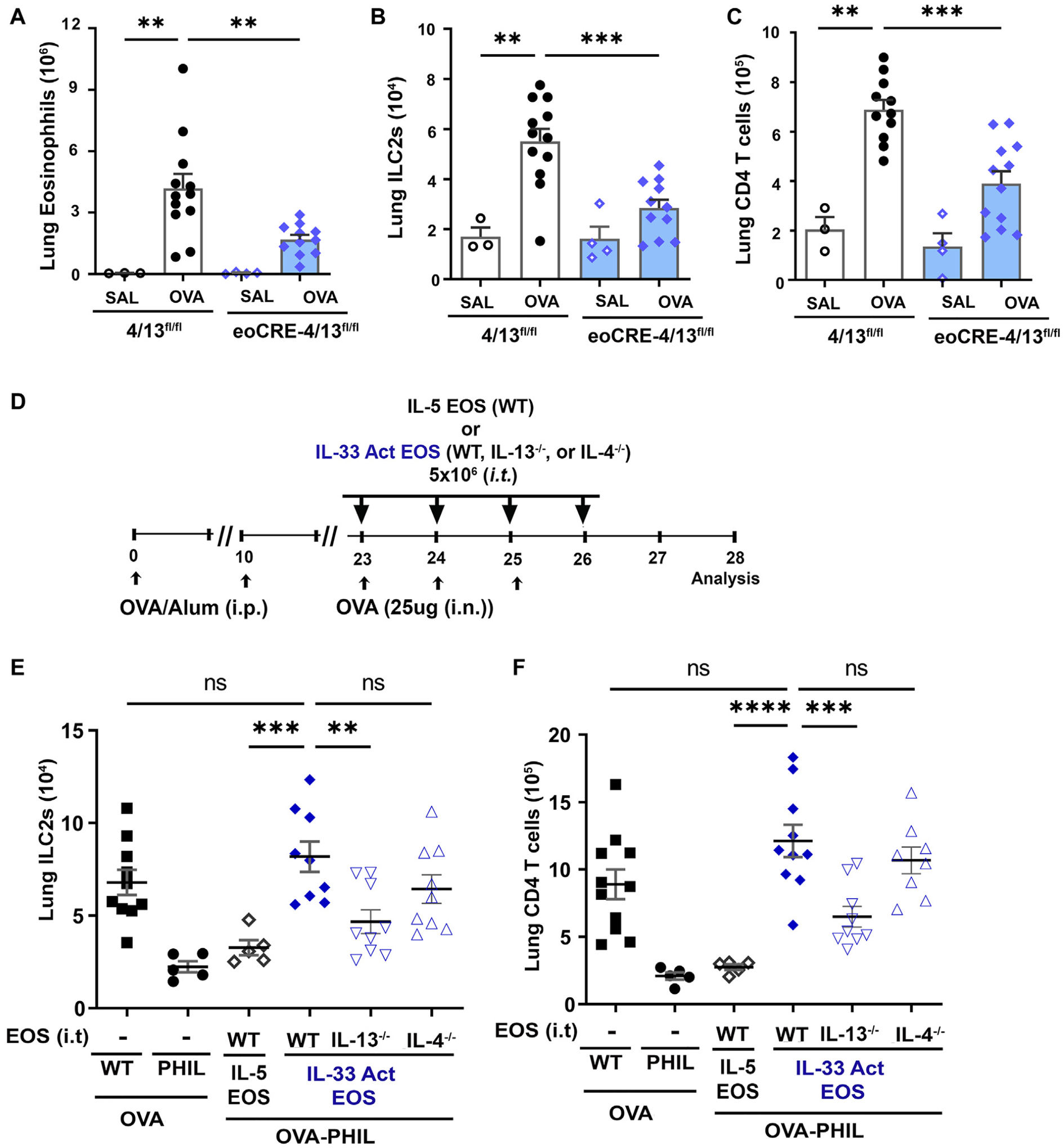
Background: Group 2 innate lymphoid cells (ILC2s) are critical mediators of type 2 respiratory inflammation, releasing IL-5 and IL-13 and promoting the pulmonary eosinophilia associated with allergen provocation. Although ILC2s have been shown to promote eosinophil activities, the role of eosinophils in group 2 innate lymphoid cell (ILC2) responses is less well defined.

Objective: We sought to investigate the role of eosinophils in activation of ILC2s in models of allergic asthma and in vitro.

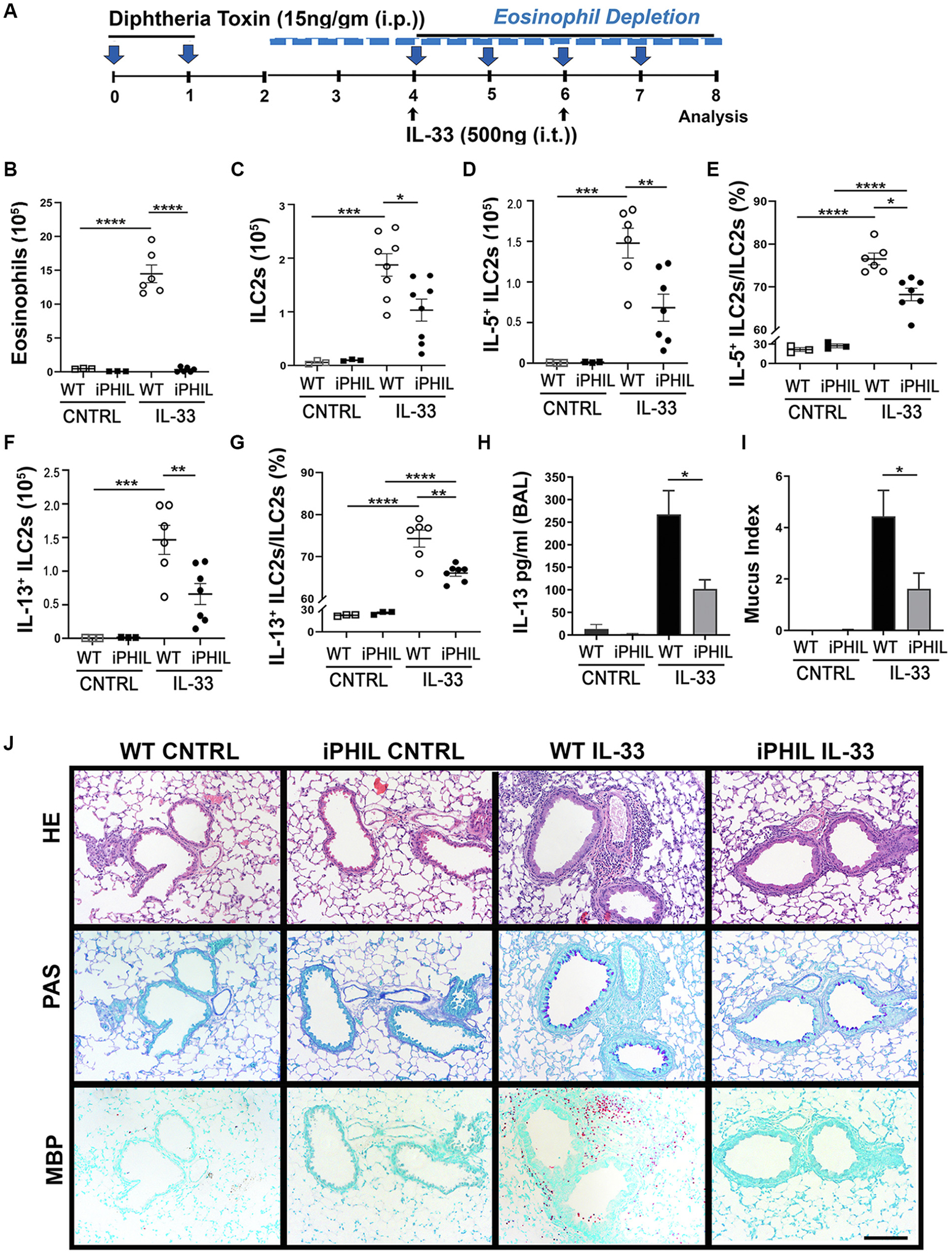
Methods: Inducible eosinophil-deficient mice were exposed to allergic respiratory inflammation models of asthma, such as ovalbumin or house dust mite challenge, or to innate models of type 2 airway inflammation, such as inhalation of IL-33. Eosinophil-specific IL-4/13-deficient mice were used to address the specific roles for eosinophil-derived cytokines. Direct cell interactions between ILC2s and eosinophils were assessed by in vitro culture experiments.

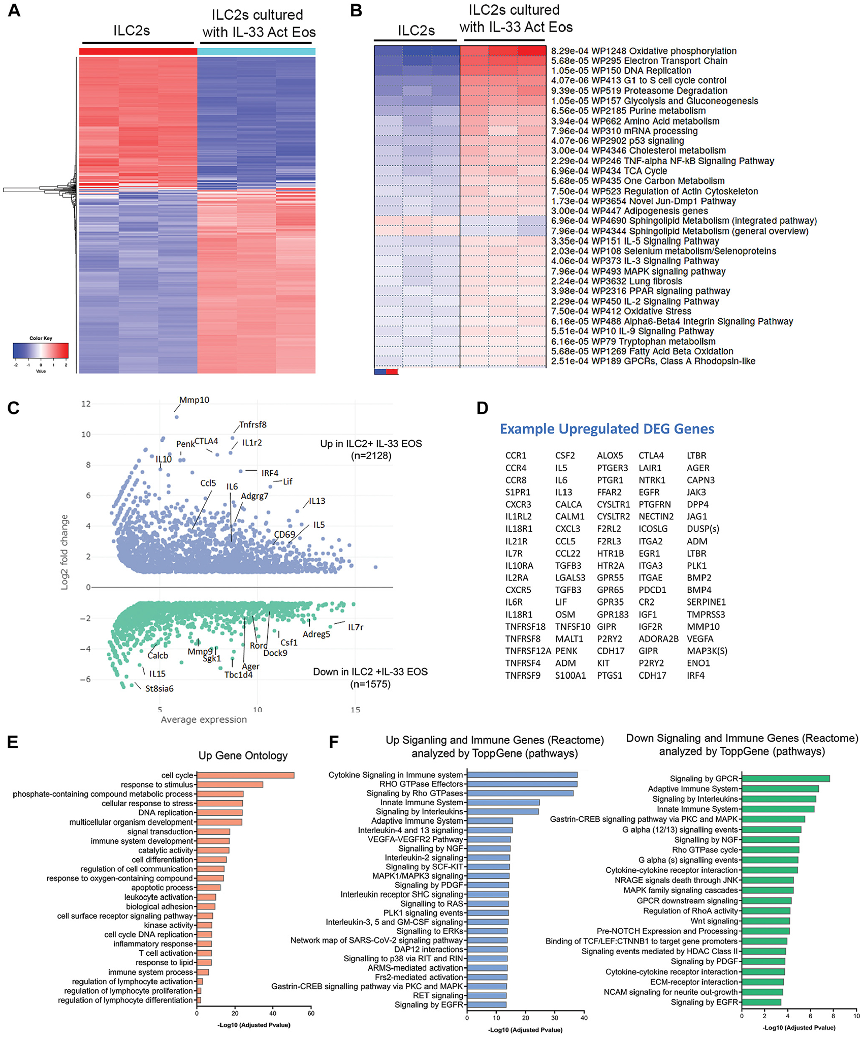
Results: Targeted depletion of eosinophils resulted in significant reductions of total and IL-5+ and IL-13+ lung ILC2s in all models of respiratory inflammation. This correlated with reductions in IL-13 levels and mucus in the airway. Eosinophil-derived IL-4/13 was necessary for both eosinophil and ILC2 accumulation in lung in allergen models. In vitro, eosinophils released soluble mediators that induced ILC2 proliferation and G protein-coupled receptor-dependent chemotaxis of ILC2s. Coculture of ILC2s and IL-33-activated eosinophils resulted in transcriptome changes in both ILC2s and eosinophils, suggesting potential novel reciprocal interactions.

Conclusion: These studies demonstrate that eosinophils play a reciprocal role in ILC2 effector functions as part of both adaptive and innate type 2 pulmonary inflammatory events.

Keywords: Eosinophil; IL-13; IL-33; IL-4; asthma; eosinophil deficient; group 2 innate lymphoid cell; inflammation; lung.


Conflict of interest statement

Disclosure of potential conflict of interest: The authors declare that they have no relevant conflicts of interest.

Figures

FIG 1.
FIG 1.
Eosinophil-depletion during OVA allergen challenge significantly reduced ILC2 accumulation and activation and type 2 pulmonary inflammation. (A) Scheme representing OVA experimental model with DT treatment in WT and eosinophil-inducible depletion iPHIL mice. Single cell lung suspensions were measured by flow cytometry. Total number of eosinophils in (B) BAL fluid and (C) lung. Total lung ILC2s (D) and total lung (E) IL-5+ ILC2s and (F) IL-13+ ILC2s. (G) Representative flow cytometry plots of ILC2s (CD45+LinCD90+Sca-1+KLRG1+) for expression of IL-5 and IL-13. Y-axis shows C90; x-axis, IL-5 or IL-13 after gating for ILC2s. Values in parentheses are cytokine-positive ILC2s out of live cells. Summary of data for percentage (H) IL-5+ ILC2s/ILC2s and (I) IL-13+ ILC2s/ILC2s in lung. (J) Total IL-13+ TH2 cells in lung. (K) BAL fluid IL-13 levels measured by ELISA. (L) Mucus index shows relative PAS-positive pixels per airway. (M) Representative images of HE-stained slides, and PAS-stained slides for mucus (purple stain) in airway. Immunohistochemistry for major basic protein 1 (MBP-1) to identify eosinophils in lung tissue. Scale bar = 100 μm. Data are shown as means 6 SEMs (n = 4–8 mice) from 2 to 3 independent experiments. Student t test (K and L) or 1-way ANOVA with Tukey multiple comparison test (B, C, D, E, G, H, I, and J). ****P < .0001, ***P < .001, **P < .01, *P < .05.
FIG 2.
FIG 2.
Eosinophil-depletion during HDM allergen challenge resulted in reduced pulmonary ILC2 accumulation and type 2 inflammation. (A) Scheme representing HDM experimental model with DT treatment in WT and iPHIL mice. Total number of eosinophils in (B) BAL fluid and (C) lung. (D) Total lung ILC2s and total lung (E) IL-5+ ILC2s and (F) IL-13+ ILC2s by intracellular flow cytometry. (G) Total IL-13+ TH2 cell in lung. (H) BAL fluid IL-13 levels measured by ELISA. (I) Mucus index shows relative PAS-positive pixels per area of airway. (J) Representative slides of lunghistologyfor HE, PAS, and immunohistochemistryfor MBP-1.Scale bar = 100 μm.Data are shown as means ± SEMs(n = 3–9mice)from 2 to 3 independent experiments by Student t test(HandI)or 1-wayANOVA with Tukey multiple comparison test (B, C, D, E, F, and G). ****P < .0001, ***P < .001, **P < .01, *P < .05.
FIG 3.
FIG 3.
Eosinophil-derived IL-4 and IL-13 contribute to network of ILC2 and T cell accumulation in an allergic model of asthma. Mice with eosinophil-specific knockout of IL-4/13 (eoCre-4/13fl/fl) and their littermate controls (4/13fl/fl) underwent OVA sensitization/challenge. Total lung (A) eosinophils, (B) ILC2s, and (C) CD4 T cells in lung were determined by flow cytometry. (D) Scheme representing OVA experimental model with intratracheal adoptive transfer of eosinophils into eosinophil-deficient PHIL mice. WT mice and PHIL mice were provided saline as controls. Cohorts of PHIL mice received IL-5–treated WT or IL-33–activated WT, IL-13−/−, or IL-4−/− eosinophils that were adoptively transferred intratracheally during allergen challenge. Total (E) ILC2s and (F) CD4 T cells in lung were determined by flow cytometry. Data are shown as means ± SEMs (n = 3–12 mice) from 3 independent experiments by 1-way ANOVA with Tukey multiple comparison test (A, B, and C). To specifically determine the significance between adoptive transfer mice, we used 1-way ANOVA with Dunnet test and control group (WT IL-33 Act EOS) (D and E). ****P < .0001, ***P < .001, **P < .01, ns, nonsignificant.
FIG 4.
FIG 4.
Eosinophils are required for IL-33–induced lung inflammation. (A) Scheme representing IL-33 experimental model with DT treatment in WT and iPHIL mice. Total lung (B) eosinophils and (C) ILC2s were determined by flow cytometry. Total (D) IL-5+ ILC2s and (E) percentage of IL-5+ ILC2s/ILC2s and total (F) IL-13+ ILC2s and (G) percentage of IL-131 ILC2s/ILC2s were measured by flow cytometry. (H) BAL fluid was measured for IL-13 by ELISA. (I) Mucus index shows relative PAS-positive pixels per area of airway. (J) Representative slides of lungs histology for HE, PAS, and immunohistochemistry for MBP-1. Scale bar = 100 μm. Data are shown as means ± SEMs (n = 3–8 mice) from 2 to 3 independent experiments by Student ttest (H and I) or 1-way ANOVA with Tukey multiple comparison test (B, C, D, E, F, and G). ****P < .0001, ***P < .001, **P < .01, *P < .05.
FIG 5.
FIG 5.
IL-33–activated eosinophils induce chemotaxis of ILC2s in a GPCR-dependent mechanism. (A) Isolated pure populations of peripheral blood eosinophils and lung-derived ILC2s were obtained to complete in vitro migration assays. Cell-free media from 48-hour cultures that included no cytokines (CN), IL-5 media, or IL-33 Act media that had been cultured with or without eosinophils were used to measure migration. After 1 hour, relative counts of ILC2s in the bottom well were counted and normalized to no cytokine in media samples. (B) Chemokinesis was tested by adding cell-free media from IL-33 Act media with eosinophils (IL-33 Act media + EOS) to either both top and bottom wells or bottom wells alone. (C) ILC2s were pretreated for 1 hour with increasing doses of PTX, then tested for response to IL-33 Act media + EOS. Migration was normalized to IL-33 Act media without EOS. Data are shown as means ± SEMs. Each data symbol represents mean value per experiment, where each experiment included >3 technical replicates. A total of 3–6 independent experiments were completed per study. We used 1-way ANOVA with Tukey multiple comparison test (A and B). To measure dose response to PTX, we used 1-way ANOVA with Dunnet test and control group IL-33 Act media + EOS 0 ng/mL PTX (C). ****P < .0001, ***P < .001. PTX, Pertussis toxin.
FIG 6.
FIG 6.
IL-33–activated eosinophils promote ILC2 activation in vitro and IL-5–treated eosinophils are sufficient to promote proliferation. Rested ILC2s were cocultured in cytokine-free media for 24 hours with no eosinophils, IL-5 EOS, or IL-33 Act EOS and then measured for morphology, cytokine expression, and proliferation at the end of 24 hours. (A) Representative HE staining of ILC2s. Scale bar = 10 μm. (B) Representative plots of intracellular IL-5 and IL-13 in ILC2s. (C) Cell-free supernatant from cocultures was measured for cytokines by multiplex assay. ILC2s were cultured alone, with IL-5 EOS, or with IL-33 Act EOS. IL-5 EOS and IL-33 Act EOS were cultured alone (no ILC2s) (right). (D) Cultures that prevented cell contact between ILC2s and eosinophils by 0.4 μm Transwell inserts (no contact) and cultures that allowed contact (contact) were measured by flow cytometry for intracellular IL-5 and IL-13 in ILC2s and (E) proliferation by Ki-67. Data are shown as means ± SEMs. One-way ANOVA with Tukey multiple comparison test (C). A representative of 3 independent experiments is shown in (D) and (E). We used 2-way ANOVA with Tukey multiple comparison test (D and E). ****P < .0001, ***P < .001, **P < .01. Specific significances are shown for ease of viewing differences between groups and conditions of “contact” and “no contact” and treatment conditions.
FIG 7.
FIG 7.
IL-33–activated eosinophils induce transcriptome changes in rested ILC2s. ILC2s and eosinophils were cultured for 24 hours as in Fig 6, then isolated for RNA-Seq. Comparisons were made between ILC2s cultured alone or with IL-33 Act EOS. (A) The 1000 most variable genes are represented in a hierarchical clustering heat map. (B) Parametric gene set enrichment (aka PGSEA) of WikiPathways. (C) MFA plot of differentially expressed genes that are FDR < 0.05 and absolute log2-fold < 1. Representative genes are labeled in MFA plot, and (D) upregulated DEG genes are shown in a table. (E) DEG genes that were FDR < 0.05 log2-fold > 1 were used to identify significant GO enrichment pathways. DEG genes that mapped to Reactome Immune System and Signal Transduction data sets were analyzed in ToppGene to identify enrichment in significant functional pathways that were both up- and downregulated between ILC2s with and without IL-33 Act EOS. MFA, Multifactor analysis.
FIG 8.
FIG 8.
ILC2s induce transcriptome changes in IL-33–activated eosinophils. ILC2s and eosinophils were cultured as in Fig 6 for 24 hours, then isolated by flow cytometry to isolate pure cell populations from culture for RNA-Seq. Comparisons were made between IL-33 Act EOS cultured with or without ILC2s. (A) Top 1000 variable genes in hierarchical clustering heat map. (B) Parametric gene set enrichment (aka PGSEA) for WikiPathways. (C) MFA plot of differentially expressed genes that are FDR < 0.05 and absolute log2-fold < 1. Representative genes are labeled in MFA plot, and (D) upregulated DEG genes are shown in a table. (E) DEG genes that were FDR < 0.05 log2-fold > 1 were used to identify significant GO-enriched pathways. DEG genes that mapped to Reactome Immune System and Signal Transduction data sets were analyzed in ToppGene to identify enrichment in significant functional pathways that were upregulated between IL-33 Act EOS with and without ILC2s. MFA, Multifactor analysis.

Similar articles

Cited by

References

    1. Hammad H, Lambrecht BN. The basic immunology of asthma. Cell 2021;184:1469–85. - PubMed
    1. Bartemes KR, Kita H. Roles of innate lymphoid cells (ILCs) in allergic diseases: the 10-year anniversary for ILC2s. J Allergy Clin Immunol 2021;147:1531–47. - PMC - PubMed
    1. Neill DR, Wong SH, Bellosi A, Flynn RJ, Daly M, Langford TK, et al. Nuocytes represent a new innate effector leukocyte that mediates type-2 immunity. Nature 2010;464(7293):1367–70. - PMC - PubMed
    1. Doherty TA, Broide DH. Airway innate lymphoid cells in the induction and regulation of allergy. Allergol Int 2019;68:9–16. - PMC - PubMed
    1. Bartemes KR, Kephart GM, Fox SJ, Kita H. Enhanced innate type 2 immune response in peripheral blood from patients with asthma. J Allergy Clin Immunol 2014;134:671–8.e4. - PMC - PubMed

Publication types