

Cdc2-like kinases: structure, biological function, and therapeutic targets for diseases
- PMID: 37029108
- PMCID: PMC10082069
- DOI: 10.1038/s41392-023-01409-4
Cdc2-like kinases: structure, biological function, and therapeutic targets for diseases
Abstract
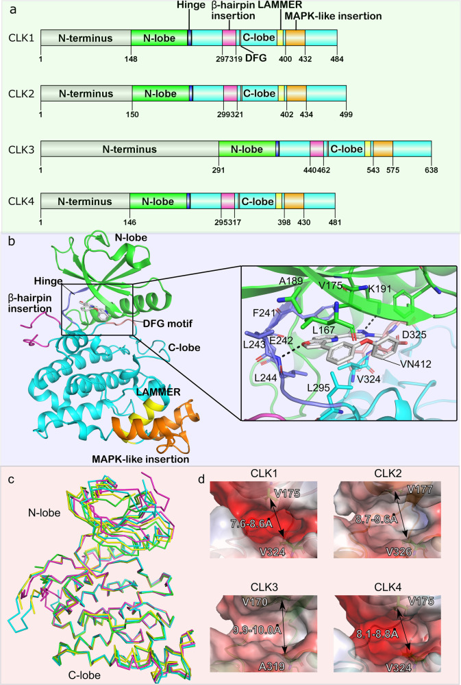
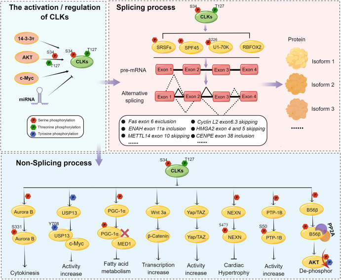
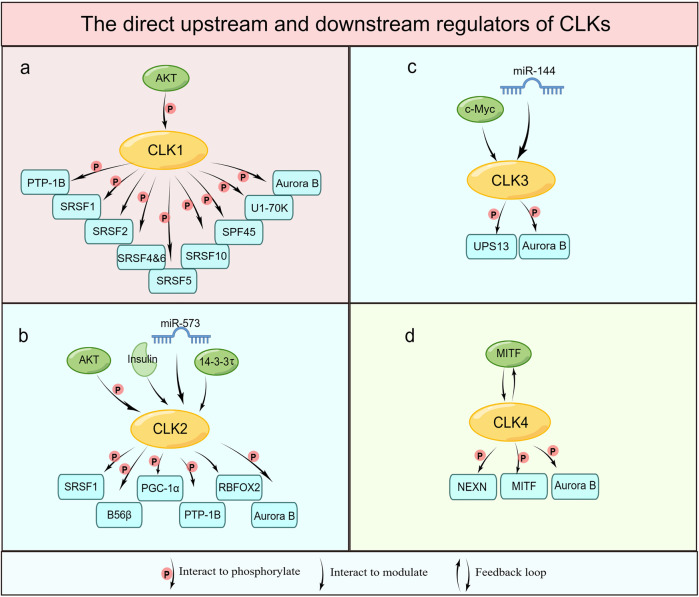
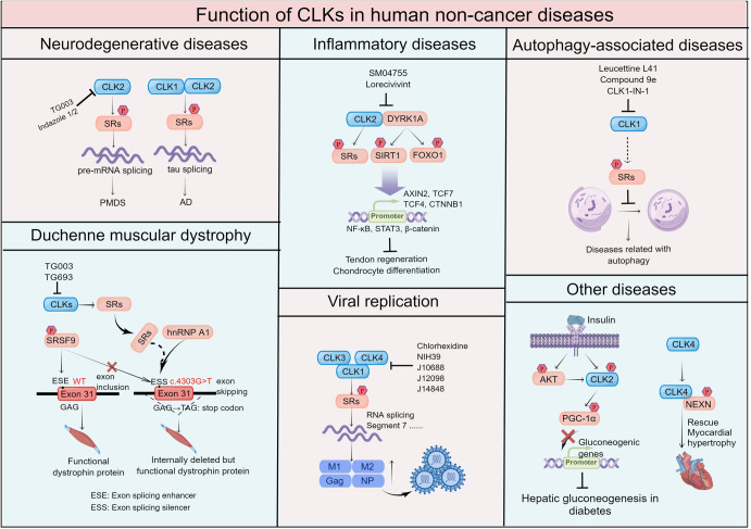
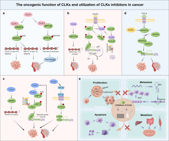
The CLKs (Cdc2-like kinases) belong to the dual-specificity protein kinase family and play crucial roles in regulating transcript splicing via the phosphorylation of SR proteins (SRSF1-12), catalyzing spliceosome molecular machinery, and modulating the activities or expression of non-splicing proteins. The dysregulation of these processes is linked with various diseases, including neurodegenerative diseases, Duchenne muscular dystrophy, inflammatory diseases, viral replication, and cancer. Thus, CLKs have been considered as potential therapeutic targets, and significant efforts have been exerted to discover potent CLKs inhibitors. In particular, clinical trials aiming to assess the activities of the small molecules Lorecivivint on knee Osteoarthritis patients, and Cirtuvivint and Silmitasertib in different advanced tumors have been investigated for therapeutic usage. In this review, we comprehensively documented the structure and biological functions of CLKs in various human diseases and summarized the significance of related inhibitors in therapeutics. Our discussion highlights the most recent CLKs research, paving the way for the clinical treatment of various human diseases.
© 2023. The Author(s).
Conflict of interest statement
The authors declare no competing interests.
Figures

Similar articles
-
Mobilization of a splicing factor through a nuclear kinase-kinase complex.Biochem J. 2018 Feb 14;475(3):677-690. doi: 10.1042/BCJ20170672. Biochem J. 2018. PMID: 29335301 Free PMC article.
-
Conserved proline-directed phosphorylation regulates SR protein conformation and splicing function.Biochem J. 2015 Mar 1;466(2):311-22. doi: 10.1042/BJ20141373. Biochem J. 2015. PMID: 25529026 Free PMC article.
-
Dual-Specificity, Tyrosine Phosphorylation-Regulated Kinases (DYRKs) and cdc2-Like Kinases (CLKs) in Human Disease, an Overview.Int J Mol Sci. 2021 Jun 3;22(11):6047. doi: 10.3390/ijms22116047. Int J Mol Sci. 2021. PMID: 34205123 Free PMC article. Review.
-
Disordered protein interactions for an ordered cellular transition: Cdc2-like kinase 1 is transported to the nucleus via its Ser-Arg protein substrate.J Biol Chem. 2019 Jun 14;294(24):9631-9641. doi: 10.1074/jbc.RA119.008463. Epub 2019 May 7. J Biol Chem. 2019. PMID: 31064840 Free PMC article.
-
A critical update on the strategies towards small molecule inhibitors targeting Serine/arginine-rich (SR) proteins and Serine/arginine-rich proteins related kinases in alternative splicing.Bioorg Med Chem. 2022 Sep 15;70:116921. doi: 10.1016/j.bmc.2022.116921. Epub 2022 Jul 9. Bioorg Med Chem. 2022. PMID: 35863237 Review.
Cited by
-
Bioinformatics analysis and experimental verification of the cancer-promoting effect of DHODH in clear cell renal cell carcinoma.Sci Rep. 2024 May 25;14(1):11985. doi: 10.1038/s41598-024-62738-0. Sci Rep. 2024. PMID: 38796629 Free PMC article.
-
Targeted therapy for knee osteoarthritis: From basic to clinics.Medicine (Baltimore). 2025 Aug 15;104(33):e43686. doi: 10.1097/MD.0000000000043686. Medicine (Baltimore). 2025. PMID: 40826764 Free PMC article. Review.
-
Small molecules modulating RNA splicing: a review of targets and future perspectives.RSC Med Chem. 2024 Jan 11;15(4):1109-1126. doi: 10.1039/d3md00685a. eCollection 2024 Apr 24. RSC Med Chem. 2024. PMID: 38665842 Free PMC article. Review.
-
Structure-guided screening identified bioactive phytoconstituents Hernandonine and Anolobine as potential inhibitors of dual specificity protein kinase CLK1.Sci Rep. 2025 Apr 19;15(1):13604. doi: 10.1038/s41598-025-97753-2. Sci Rep. 2025. PMID: 40253440 Free PMC article.
-
Identification and Biological Evaluation of a Novel CLK4 Inhibitor Targeting Alternative Splicing in Pancreatic Cancer Using Structure-Based Virtual Screening.Adv Sci (Weinh). 2025 May;12(19):e2416323. doi: 10.1002/advs.202416323. Epub 2025 Mar 24. Adv Sci (Weinh). 2025. PMID: 40126184 Free PMC article.
References
Publication types
MeSH terms
Substances
LinkOut - more resources
Full Text Sources
Research Materials
Miscellaneous
