. 2023 Apr 7;8(1):52.
doi: 10.1038/s41541-023-00653-7.

Glycosylated nanoparticle-based PfCSP vaccine confers long-lasting antibody responses and sterile protection in mouse malaria model

Affiliations

Glycosylated nanoparticle-based PfCSP vaccine confers long-lasting antibody responses and sterile protection in mouse malaria model

Julia Ludwig et al. NPJ Vaccines. .

Erratum in

Abstract

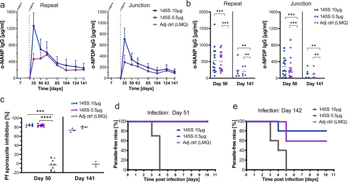
The development of an effective and durable vaccine remains a central goal in the fight against malaria. Circumsporozoite protein (CSP) is the major surface protein of sporozoites and the target of the only licensed Plasmodium falciparum (Pf) malaria vaccine, RTS,S/AS01. However, vaccine efficacy is low and short-lived, highlighting the need for a second-generation vaccine with superior efficacy and durability. Here, we report a Helicobacter pylori apoferritin-based nanoparticle immunogen that elicits strong B cell responses against PfCSP epitopes that are targeted by the most potent human monoclonal antibodies. Glycan engineering of the scaffold and fusion of an exogenous T cell epitope enhanced the anti-PfCSP B cell response eliciting strong, long-lived and protective humoral immunity in mice. Our study highlights the power of rational vaccine design to generate a highly efficacious second-generation anti-infective malaria vaccine candidate and provides the basis for its further development.


Conflict of interest statement

S.H., J. Lossin, A.O., N.L., B.F., C.J.J., S. B., P.K. declare no conflicts of interest. J. Ludwig, S.W.S., G.C., R.M., K.P., H.W., E.A.L. and J.-P.J. have filed a patent application related to the immunogens described in this work.

Figures

Fig. 1
Fig. 1. The C-CSP of recombinant PfCSP is highly immunogenic but fails to induce protective serum-antibody responses in mice.
a C57BL/6J mice were immunized with FL-PfCSP at day 0, 21 and 42 adjuvanted with SAS. The serum IgG concentration against the indicated PfCSP domains was determined 7 days after the last immunization. Dots represent data from individual mice. Pooled data of three independent experiments with 5 mice per group are shown. Black horizontal lines indicate arithmetic means. Statistically significant differences were calculated by Kruskal–Wallis test with Dunn’s correction (*P < 0.05; **P < 0.01; ****P < 0.0001). b Sera, collected 7 days after the last immunization, from 5 mice of the same experimental group were pooled and NANP-reactive antibodies were depleted. The IgG concentration of NANP and C-CSP reactive antibodies was determined before (pre) and after (post) depletion. Dots represent independent experiments. c Binding profile to Pf sporozoites of the pooled sera described in (b) diluted 1:100. Gating on Pf sporozoites is shown in Supplementary Fig. 1. d Capacity of the pooled sera described in (c) to inhibit the hepatocyte traversal activity of Pf sporozoites in vitro. Dots represent independent traversal assay experiments. Gating strategy for Pf traversal analysis is shown in Supplementary Fig. 2.
Fig. 2
Fig. 2. Design and immunogenicity of a PfCSP nanoparticle immunogen.
a Schematic representation of immunogen 126, comprising the PfCSP epitope motifs KQPADG (brown), NPDP (green), NANP (blue) and NVDP (yellow) genetically fused to H. pylori apoferritin (gray) and PADRE (pink), separated by short linkers (black). Models of immunogen 126 displayed as both a monomer and as an assembled nanoparticle. The PfCSP epitope is presented externally on the nanoparticle surface. For better illustration, six monomers have been removed from the assembled nanoparticle, thus slicing through the immunogen, to reveal the PADRE epitopes within its core. b Size exclusion chromatogram and SDS-PAGE analysis of immunogen 126 (uncropped SDS-PAGE in Supplementary Fig. 3a). c Negative stain electron microscopy of immunogen 126. Scale bar—50 nm. d C57BL/6J mice were immunized with FL-PfCSP or immunogen 126 adjuvanted with SAS at day 0, 21 and 42. The serum IgG response against the PfCSP repeat (NANP) and junction (NPDP) was determined at the indicated timepoints. Immunization with SAS alone served as negative control. Pooled data of two independent experiments with 5 mice per group are shown. e Mice from the Kymouse™ platform were immunized with FL-PfCSP or immunogen 126 adjuvanted with SAS at day 0, 28 and 70. The serum IgG response against the PfCSP repeat (NANP) and the junction (NPDP) was measured at the indicated timepoints. Immunization with SAS alone served as control. One representative out of two independent experiments with 7 mice per group is shown. f IGHV3-33 (left) and paired IGHV3-33/IGKV1-5 (right) gene usage frequency among sorted GC CSP+ or GC CSP− cells isolated from lymph nodes of mice from the Kymouse™ platform 7 days after the third immunization with immunogen 126 adjuvanted with SAS. Data from naïve B cells are shown for comparison. FACS gating strategy is shown in Supplementary Fig. 6. Dots represent individual mice. Pooled data of two independent experiments are shown (CSP−: n = 6, CSP+: n = 4; naïve: n = 20). g Silent (S, gray) and replacement (red) mutations in VH3-33 antibodies of mice from the Kymouse™ platform. CDRs are marked in light gray. Arrows indicate positions H.31 and H.50 with strong selection for replacement mutations. h Capacity of pooled sera (diluted 1:400, collected 7 days after the last immunization) from mice of the Kymouse™ platform to inhibit the hepatocyte traversal activity of Pf sporozoites in vitro. Dots represent independent traversal assay experiments (n = 4). Arithmetic mean (d, e, h), median with length of the whiskers as multiple of IQR (f) and SEM (d, e) are indicated. Statistically significant differences were calculated by two-tailed Mann–Whitney test. In (d) and (e) statistical analyses were performed with data from day 50 and day 80, respectively (*P < 0.05; **P < 0.01; ***P < 0.001). Statistically non-significant differences are not indicated.
Fig. 3
Fig. 3. Glycan modifications of the nanoparticle focus the humoral response on PfCSP epitopes.
a Schematic representation of immunogen 145S. Immunogen 126 (Fig. 2a) was modified by the engineering of two non-native N-linked glycosylation sites at positions N79 and N99 of H. pylori apoferritin. Expression of these nanoparticles in HEK293S (GnT I−/−) cells led to an addition of high mannose sugars at the targeted positions. This glycosylation (colored light green) covers the H. pylori apoferritin nanoparticle surface while not affecting presentation of the PfCSP epitope (scheme below). b Size exclusion chromatogram and SDS-PAGE analysis of immunogen 145S (uncropped SDS-PAGE in Supplementary Fig. 3b). c Negative stain electron microscopy of immunogen 145S. Scale bar—50 nm. d C57BL/6J mice were immunized with immunogen 126 or 145S adjuvanted with LMQ at day 0 and 28. The serum IgG response against H. pylori apoferritin was measured at various timepoints. Immunization with LMQ alone served as control. One representative out of two independent experiments with 5 mice per group is shown. e H. pylori apoferritin IgG response 22 days after the last immunization with immunogen 145S or 126 compared to adjuvant alone as negative control. Dots represent individual mice. Pooled data of two independent experiments with 5 mice per group are shown. Symbols indicate independent experiments. f Serum IgG response of the mice described in (d) against the PfCSP repeat (NANP) and the junction (NPDP). One representative out of two independent experiments with 5 mice per group is shown. g Comparison of the IgG response against the PfCSP repeat (NANP) and junction (NPDP) at 7 days and 22 days after the last immunization. Dots represent individual mice. Pooled data of two independent experiments with 5 mice per group are shown. Symbols indicate independent experiments. h Representative FACS analysis (left) and quantification (right) of live IgG+ antigen-binding cells in lymph nodes of mice immunized with immunogen 126, 145S or adjuvant alone 22 days after the last immunization. Dots represent individual mice. Pooled data of two independent experiments are shown. Symbols indicate independent experiments. Dark red (n = 6) and blue (n = 9): 10 µg dose immunization; light red (n = 5) and violet (n = 5): 0.5 µg dose immunization; adjuvant control group (n = 5). Detailed gating strategy is shown in Supplementary Fig. 9. i ELISpot-based enumeration of H. pylori apoferritin and FL-CSP reactive bone marrow plasma cells (PCs) of mice 22 days after the last immunization with immunogen 145S or 126 at 10 µg (dark red (n = 6) and blue (n = 9), respectively) or 0.5 µg (light red (n = 5) and violet (n = 5), respectively) or adjuvant alone (n = 10). Dots represent individual mice. Pooled data of two independent experiments are shown. Symbols indicate independent experiments. j Capacity of pooled sera (diluted 1:800, collected 22 days after the last immunization) from the same mice as in (i) to inhibit the hepatocyte traversal activity of Pf sporozoites in vitro. Pooled data of three independent traversal assay experiments are shown. Dark red (n = 6) and blue (n = 9): 10 µg dose; light red (n = 6) and violet (n = 6): 0.5 µg dose; adjuvant control group (n = 9). Symbols indicate independent immunization experiments. Arithmetic mean (dj) and SEM (d, f) are indicated. Statistically significant differences were calculated by two-tailed Mann–Whitney test (*P < 0.05; **P < 0.01; ***P < 0.001, ****P < 0.0001).
Fig. 4
Fig. 4. Efficient T cell help by the universal T cell epitope PADRE.
C57BL/6J mice were immunized with immunogen 145S adjuvanted with LMQ at day 0 and 28 or adjuvant alone. Representative FACS plots (left) and quantification (right) of CD4 EM cells (a), EM Tfh cells (b) and PADRE-specific CD4 T cells (c) in lymph nodes 22 days after the last immunization. Detailed gating strategy is shown in Supplementary Fig. 10. Pooled data of three independent experiments for the immunogen 145S group and two independent experiments for the adjuvant control group with 5 mice per group are shown. Dots represent individual mice. Symbols indicate independent experiments. d In vitro CFSE proliferation assay with splenocytes isolated 22 days after the last immunization. Splenocytes were restimulated with 10 µg/ml (7.4 µM) PADRE or OVA control peptide and the proportion of CFSElow cells was determined 3 days later. Representative FACS plots of EM T cells (left) after stimulation with the negative control OVA peptide (top) or PADRE (bottom). The proportion of proliferating cells was determined in different T cell populations including all gated PADRE+ CD4 T cells (right). Detailed gating strategy is shown in Supplementary Fig. 11. Pooled data of two independent experiments with two replicates for each experiment and 4 mice per group are shown. e The proportion of proliferating CD4 T cells 3 days after restimulation of splenocytes isolated 22 days after the last immunization with different concentrations of the PADRE peptide. Pooled data of two independent experiments with two replicates for each experiment and 4 mice per group are shown. Arithmetic mean (ae) and SEM (d, e) are indicated. Statistically significant differences were calculated by two-tailed Mann–Whitney test (*P < 0.05; **P < 0.01; ***P < 0.001, ****P < 0.0001, n.s. statistically non-significant differences).
Fig. 5
Fig. 5. The glycosylated nanoparticle 145S confers long-lived protective antibody responses.
a C57BL/6J mice were immunized with 10 µg or 0.5 µg immunogen 145S adjuvanted with LMQ or with LMQ alone at day 0 and 28. The serum IgG response against the repeat (NANP) and the junction (NPDP) was measured at different timepoints. One representative out of two independent experiments with 10 mice per group is shown until day 50. Day 51 until day 141 shows data from one experiment with 5 mice per group. b Anti-repeat (NANP) or junction (NPDP) IgG response at day 50 and day 141 after the first immunization. Dots represent individual mice. Day 50: Pooled data of three independent experiments with 5 mice per group are shown. Day 141: Data of one experiment with 5 mice per group is shown. Symbols indicate independent experiments. c Capacity of pooled sera (diluted 1:800) collected 50 days or 141 days after first immunization to inhibit the hepatocyte traversal activity of Pf sporozoites in vitro. Pooled data of three independent traversal assay experiments are shown. Symbols indicate independent immunization experiments. d, e Capacity of immunization with immunogen 145S at two different doses to protect mice after PbPfCSP(mCherry)-infected mosquito bite challenge (3 bites per mouse) from parasitemia. Data show the percentage of blood stage parasite-free mice over time. d Day of challenge 51: Pooled data of two independent experiments with 5 mice per group are shown. e Day of challenge 142: Data from one experiment with 5 mice per group. Arithmetic mean (ac) and SEM (a) are indicated. Statistically-significant differences were calculated by two-tailed Mann–Whitney test (*P < 0.05; **P < 0.01; ***P < 0.001, ****P < 0.0001). Statistically non-significant differences are not indicated.

References

    1. WHO. World Malaria Report 2021 (WHO Press, 2021).
    1. Sidjanski SP, Vanderberg JP, Sinnis P. Anopheles stephensi salivary glands bear receptors for region I of the circumsporozoite protein of Plasmodium falciparum. Mol. Biochem. Parasitol. 1997;90:33–41. doi: 10.1016/S0166-6851(97)00124-2. - DOI - PMC - PubMed
    1. Ménard R, et al. Circumsporozoite protein is required for development of malaria sporozoites in mosquitoes. Nature. 1997;385:336–339. doi: 10.1038/385336a0. - DOI - PubMed
    1. Frevert U, et al. Intravital observation of plasmodium berghei sporozoite infection of the liver. PLoS Biol. 2005;3:e192. doi: 10.1371/journal.pbio.0030192. - DOI - PMC - PubMed
    1. Cerami C, et al. The basolateral domain of the hepatocyte plasma membrane bears receptors for the circumsporozoite protein of plasmodium falciparum sporozoites. Cell. 1992;70:1021–1033. doi: 10.1016/0092-8674(92)90251-7. - DOI - PubMed