. 2023 Jun;22(6):e13834.
doi: 10.1111/acel.13834. Epub 2023 Apr 7.

Parkin regulates microglial NLRP3 and represses neurodegeneration in Parkinson's disease

Affiliations

Parkin regulates microglial NLRP3 and represses neurodegeneration in Parkinson's disease

Yi-Qun Yan et al. Aging Cell. 2023 Jun.

Abstract

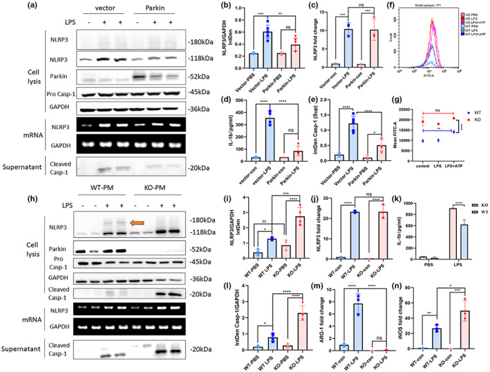
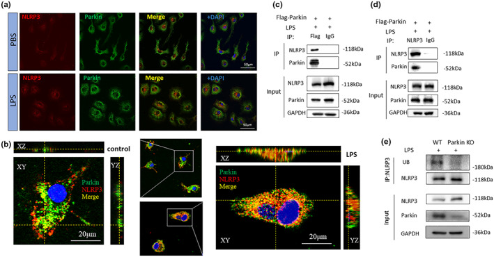
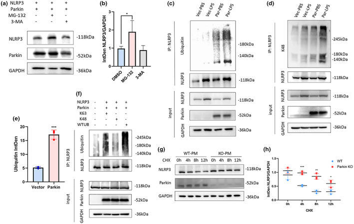
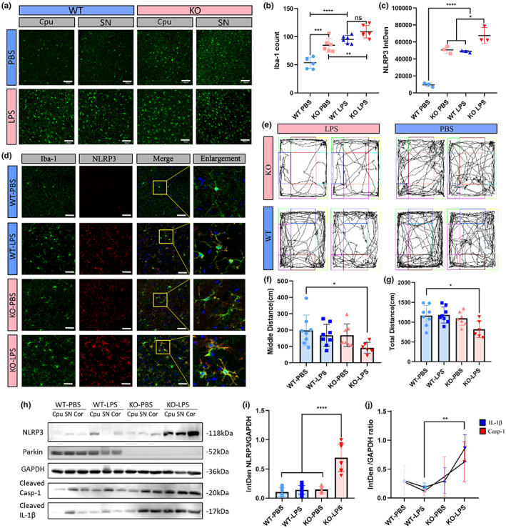
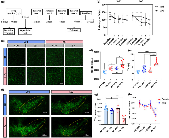
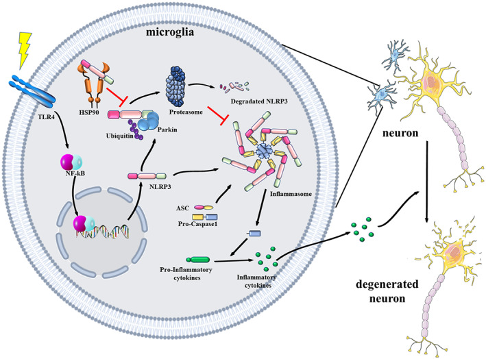
Microglial hyperactivation of the NOD-, LRR-, and pyrin domain-containing 3 (NLRP3) inflammasome contributes to the pathogenesis of Parkinson's disease (PD). Recently, neuronally expressed NLRP3 was demonstrated to be a Parkin polyubiquitination substrate and a driver of neurodegeneration in PD. However, the role of Parkin in NLRP3 inflammasome activation in microglia remains unclear. Thus, we aimed to investigate whether Parkin regulates NLRP3 in microglia. We investigated the role of Parkin in NLRP3 inflammasome activation through the overexpression of Parkin in BV2 microglial cells and knockout of Parkin in primary microglia after lipopolysaccharide (LPS) treatment. Immunoprecipitation experiments were conducted to quantify the ubiquitination levels of NLRP3 under various conditions and to assess the interaction between Parkin and NLRP3. In vivo experiments were conducted by administering intraperitoneal injections of LPS in wild-type and Parkin knockout mice. The Rotarod test, pole test, and open field test were performed to evaluate motor functions. Immunofluorescence was performed for pathological detection of key proteins. Overexpression of Parkin mediated NLRP3 degradation via K48-linked polyubiquitination in microglia. The loss of Parkin activity in LPS-induced mice resulted in excessive microglial NLRP3 inflammasome assembly, facilitating motor impairment, and dopaminergic neuron loss in the substantia nigra. Accelerating Parkin-induced NLRP3 degradation by administration of a heat shock protein (HSP90) inhibitor reduced the inflammatory response. Parkin regulates microglial NLRP3 inflammasome activation through polyubiquitination and alleviates neurodegeneration in PD. These results suggest that targeting Parkin-mediated microglial NLRP3 inflammasome activity could be a potential therapeutic strategy for PD.

Keywords: NLRP3; Parkin; microglia; neuroinflammation; ubiquitination.


Conflict of interest statement

All authors claim that there are no conflicts of interest.

Figures

FIGURE 1
FIGURE 1
Parkin regulates NLRP3 inflammasome activation. (a) Overexpression of Parkin in BV2 cells alleviates NLRP3 inflammasome activation and the downstream inflammatory response but does not change the NLRP3 mRNA level. (b, i) Statistical analysis of the NLRP3 integrated density of WB experiments in BV2 cells (b) and PM (i). (d, k) ELISA of IL‐1β in the supernatant of BV2 cells (d) and PM (k). (c, j) qPCR of NLRP3 in BV2 (c) and PM (j). (e) Statistical analysis of cleaved Caspase‐1levels in supernatant of BV2. (f) Mitochondrial ROS levels of WT and Parkin KO PM quantified by flow cytometric analysis. (g) Statistical analysis of mean FTIC‐A in flow cytometric experiments. (h) Parkin KO exacerbates NLRP3 inflammasome activation and the downstream inflammatory response but does not alter the NLRP3 mRNA level. (l) Statistical analysis of the cleaved Caspase‐1 integrated density of WB experiments in PM. (m, n) qPCR of ARG‐1, and iNOS in PM. The orange arrow points out the post‐translational band of NLRP3. NLRP3, NOD‐, LRR‐, and pyrin domain‐containing 3; WB, Western blot; PM, primary microglia; ELISA, enzyme‐linked immunosorbent assay; IL‐1β, interleukin 1 beta; qPCR, quantitative polymerase chain reaction; ROS, reactive oxygen species; WT, wild‐type; KO, knockout; ARG‐1, arginase 1; iNOS, inducible nitric oxide synthase; ns, not significant. *p < 0.05, **p < 0.01, ***p < 0.001, ****p < 0.0001.
FIGURE 2
FIGURE 2
Interaction between NLRP3 and Parkin. (a) Co‐localization of NLRP3 with Parkin assessed via confocal microscopy. (b) 3D‐reconstructed image of NLRP3 and Parkin in BV2 cells via two‐photon microscopy. (c, d) Co‐immunoprecipitation experiment conducted on BV2 cells using a FLAG‐Parkin antibody and an NLRP3 antibody. (e) Immunoprecipitation conducted on WT or Parkin KO primary microglia using NLRP3 antibody. Ubiquitin and NLRP3 was detected. NLRP3, NOD‐, LRR‐ and pyrin domain‐containing 3; WT, wield type; KO, knockout.
FIGURE 3
FIGURE 3
(a, b) Influence of two inhibitors, MG‐132, and 3‐MA on 293 T cells transfected with a Parkin and NLRP3 expression plasmid (a). Statistical analysis of NLRP3 levels in MG‐132, 3‐MA groups (b). (c–e) IP of NLRP3 in BV2 cells. Ubiquitination levels measured using a ubiquitin antibody (c) and a K48‐linked ubiquitin antibody (d). Statistical analysis of anti‐ubiquitin bands (e). (f) IP of NLRP3 in 293 T cells transfected with a different ubiquitin plasmid. (g, h) cycloheximide chase experiment on WT and Parkin KO primary microglia. Degradation speed was faster in WT group than in Parkin KO group (g), Statistical analysis showed difference at 4, 8, and 12 h time points (h). 3‐MA, 3‐methyl adenine; IP, immunoprecipitation; KO, knockout; NLRP3, NOD‐, LRR‐ and pyrin domain‐containing 3; WT, wild type. *p < 0.05, **p < 0.01, ***p < 0.001.
FIGURE 4
FIGURE 4
Parkin deficiency induced stronger microglia activation and heavier inflammatory response in mice. 12 WT and 12 Parkin KO male mice were divided into four groups (n = 6): WT‐PBS, WT‐ LPS, KO‐PBS, and KO‐LPS. In each group, three were used for immunofluorescence and three were used for WB. (a, b) Iba‐1 labeling of brain sections from SNc and striatum regions and Iba‐1‐positive cell counts. Each dot represents the SNc or striatum region of one mouse. (c, d) NLRP3 and Iba‐1 co‐labeling of brain sections of mice and statistical analysis of the integrated density of NLRP3 levels. Each dot represents one mouse. (e–g) Motion trajectories of mice (e) and distance traveled in the whole open field (f) or the middle area of the open field apparatus (g). (h–j) WB of NLRP3 inflammatory proteins in the brains of mice. Scale bar: 50 μm (a), 30 μm (d). KO, knock; LPS, lipopolysaccharide; NLRP3, NOD‐, LRR‐, and pyrin domain‐containing 3; SNc, substantia nigra pars compacta; WB, Western blot; WT, wield type. *p < 0.05, **p < 0.01, ***p < 0.001, ****p < 0.0001.
FIGURE 5
FIGURE 5
Neuroinflammation and PD‐related pathology 6 months after LPS treatment. 16 WT (8 males and 8 females) and 16 Parkin KO (8 males and 8 females) mice were divided into four groups (equal sex distribution): WT‐PBS, WT‐ LPS, KO‐PBS, KO‐LPS. Two in KO‐LPS group and one in KO‐PBS group died during the experiment and were excluded. (a) Schematic representation illustrating the experimental design (timeline). (b) Latency to fall in the Rotarod test at 6 months. (c, d) CD11b staining of brain sections of mice and integrated density of CD11b levels. Each dot represents the SNc or striatum region of one mouse. Scale bar: 100 μm. (e) Time required to reach the bottom of the pole from the top at 6 months post‐LPS injection. (f–h) TH staining in the SNc of the brains of mice. The yellow circle indicates the SNc. Quantification of the relative number of TH‐positive cells in the SNc, as determined for the whole group (g) and separately for each sex (h), although the results are the same. Each dot represents a mouse. Scale bar: 100 μm (c), 500 μm (f). KO, knockout; LPS, lipopolysaccharide; PD, Parkinson''s disease; SNc, substantia nigra pars compacta; TH, tyrosine hydroxylase; WT, wield type. *p < 0.05, **p < 0.01, ***p < 0.001, ****p < 0.0001.
FIGURE 6
FIGURE 6
Schematic diagram of the regulatory mechanism through which Parkin and HSP90 modulate NLRP3‐associated inflammation. Microglia receive stimulation through the activation of surface receptors and then express NLRP3 and pro‐inflammatory proteins. NLRP3 can either assemble into an inflammasome or degrade through the ubiquitin‐proteasome pathway. Parkin is responsible for NLRP3 degradation by mediating K48‐linked polyubiquitination. The combination of NLRP3 with HSP90 could prevent NLRP3 from becoming degraded. NLRP3 inflammasome activation in microglia could create a cytotoxic environment for neurons and lead to neurodegeneration in Parkinson's disease. HSP90, heat shock protein 90; NLRP3, NOD‐, LRR‐ and pyrin domain‐containing 3.

Similar articles

Cited by

References

    1. Avagliano, C. , Coretti, L. , Lama, A. , Pirozzi, C. , de Caro, C. , de Biase, D. , Turco, L. , Mollica, M. P. , Paciello, O. , Calignano, A. , Meli, R. , Lembo, F. , & Mattace Raso, G. (2022). Dual‐hit model of Parkinson's disease: Impact of Dysbiosis on 6‐Hydroxydopamine‐insulted mice‐neuroprotective and anti‐inflammatory effects of butyrate. International Journal of Molecular Sciences, 23(12), 6367. 10.3390/ijms23126367 - DOI - PMC - PubMed
    1. Azambuja, J. H. , Mancuso, R. I. , Via, F. I. D. , Torello, C. O. , & Saad, S. T. O. (2022). Protective effect of green tea and epigallocatechin‐3‐gallate in a LPS‐induced systemic inflammation model. The Journal of Nutritional Biochemistry, 101, 108920. 10.1016/j.jnutbio.2021.108920 - DOI - PubMed
    1. Balestrino, R. , & Schapira, A. H. V. (2020). Parkinson disease. European Journal of Neurology, 27(1), 27–42. 10.1111/ene.14108 - DOI - PubMed
    1. Bohush, A. , Bieganowski, P. , & Filipek, A. (2019). Hsp90 and its co‐chaperones in neurodegenerative diseases. International Journal of Molecular Sciences, 20(20), E4976. Retrieved from. 10.3390/ijms20204976 - DOI - PMC - PubMed
    1. Burmann, B. M. , Gerez, J. A. , Matečko‐Burmann, I. , Campioni, S. , Kumari, P. , Ghosh, D. , Mazur, A. , Aspholm, E. E. , Šulskis, D. , Wawrzyniuk, M. , Bock, T. , Schmidt, A. , Rüdiger, S. G. D. , Riek, R. , & Hiller, S. (2020). Regulation of α‐synuclein by chaperones in mammalian cells. Nature, 577(7788), 127–132. 10.1038/s41586-019-1808-9 - DOI - PMC - PubMed

Publication types

MeSH terms

Substances

LinkOut - more resources