. 2023 Jul;165(1):71-87.
doi: 10.1053/j.gastro.2023.03.229. Epub 2023 Apr 6.

The Long Noncoding RNA Cardiac Mesoderm Enhancer-Associated Noncoding RNA (Carmn) Is a Critical Regulator of Gastrointestinal Smooth Muscle Contractile Function and Motility

Affiliations

The Long Noncoding RNA Cardiac Mesoderm Enhancer-Associated Noncoding RNA (Carmn) Is a Critical Regulator of Gastrointestinal Smooth Muscle Contractile Function and Motility

Xiangqin He et al. Gastroenterology. 2023 Jul.

Abstract

Background & aims: Visceral smooth muscle cells (SMCs) are an integral component of the gastrointestinal (GI) tract that regulate GI motility. SMC contraction is regulated by posttranslational signaling and the state of differentiation. Impaired SMC contraction is associated with significant morbidity and mortality, but the mechanisms regulating SMC-specific contractile gene expression, including the role of long noncoding RNAs (lncRNAs), remain largely unexplored. Herein, we reveal a critical role of Carmn (cardiac mesoderm enhancer-associated noncoding RNA), an SMC-specific lncRNA, in regulating visceral SMC phenotype and contractility of the GI tract.

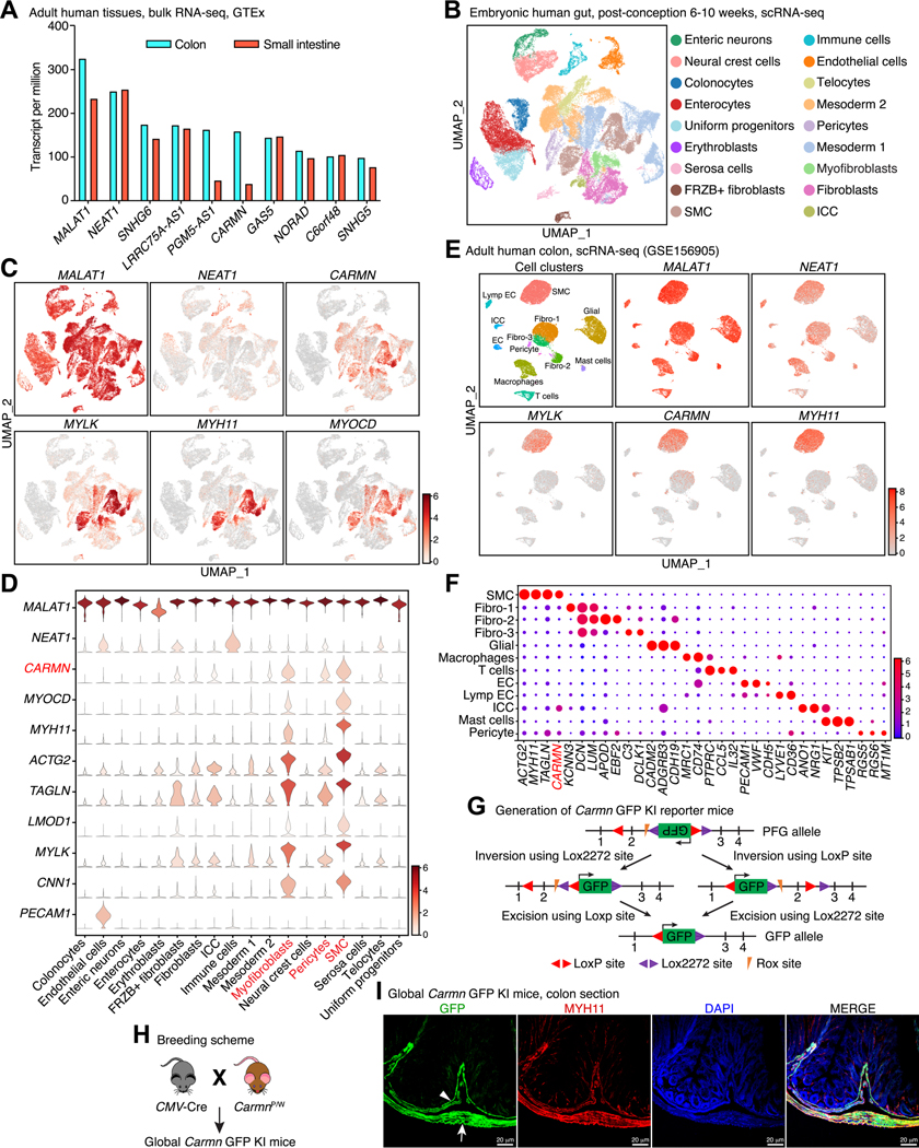
Methods: Genotype-Tissue Expression and publicly available single-cell RNA sequencing (scRNA-seq) data sets from embryonic, adult human, and mouse GI tissues were interrogated to identify SMC-specific lncRNAs. The functional role of Carmn was investigated using novel green fluorescent protein (GFP) knock-in (KI) reporter/knock-out (KO) mice. Bulk RNA-seq and single nucleus RNA sequencing (snRNA-seq) of colonic muscularis were used to investigate underlying mechanisms.

Results: Unbiased in silico analyses and GFP expression patterns in Carmn GFP KI mice revealed that Carmn is highly expressed in GI SMCs in humans and mice. Premature lethality was observed in global Carmn KO and inducible SMC-specific KO mice due to GI pseudo-obstruction and severe distension of the GI tract, with dysmotility in cecum and colon segments. Histology, GI transit, and muscle myography analysis revealed severe dilation, significantly delayed GI transit, and impaired GI contractility in Carmn KO vs control mice. Bulk RNA-seq of GI muscularis revealed that loss of Carmn promotes SMC phenotypic switching, as evidenced by up-regulation of extracellular matrix genes and down-regulation of SMC contractile genes, including Mylk, a key regulator of SMC contraction. snRNA-seq further revealed SMC Carmn KO not only compromised myogenic motility by reducing contractile gene expression but also impaired neurogenic motility by disrupting cell-cell connectivity in the colonic muscularis. These findings may have translational significance, because silencing CARMN in human colonic SMCs significantly attenuated contractile gene expression, including MYLK, and decreased SMC contractility. Luciferase reporter assays showed that CARMN enhances the transactivation activity of the master regulator of SMC contractile phenotype, myocardin, thereby maintaining the GI SMC myogenic program.

Conclusions: Our data suggest that Carmn is indispensable for maintaining GI SMC contractile function in mice and that loss of function of CARMN may contribute to human visceral myopathy. To our knowledge this is the first study showing an essential role of lncRNA in the regulation of visceral SMC phenotype.

Keywords: Carmn; Contractility; Pseudo-obstruction; Smooth Muscle; Visceral Myopathy; lncRNA.


Conflict of interest statement

Conflict of interest statement: The authors have no conflicts of interest to disclose.

Figures

Figure 1.
Figure 1.. Identification of CARMN as a SMC-specific lncRNA in human and mouse GI tissues.
(A) Analysis of the GTEx database showing the top 10 most abundant lncRNAs in human colon and intestinal tissues. (B) UMAP plot of cell populations based on scRNA-seq analysis of developing human gut tissues including duodenum, jejunum, ileum and colon. (C) UMAP visualization of the distribution of gene expression for NEAT1, CARMN, MYLK, MYH11 and MYOCD in embryonic human gut based on scRNA-seq analysis. (D) Violin plot showing the expression of selected genes in the different cell types identified in embryonic human gut. CARMN is found to be specifically expressed in SMCs, myofibroblasts and pericytes (in red). (E) UMAP plot showing cell types and the expression of selected genes in adult human colon as revealed by scRNA-seq analysis. (F) Dot plot showing the marker genes used to identify the cell types that comprise the adult human colon. (G) Strategy used to generate Carmn GFP KI mice. (H) Breeding strategy used to generate global Carmn GFP KI reporter mice. (I) Direct visualization of GFP and immunostaining of MYH11 to determine the cellular distribution of Carmn in relation to SMCs in colon dissected from K/W mice. Arrow and arrowhead denote muscularis externa SMCs and mucosa muscularis SMCs, respectively.
Figure 2.
Figure 2.. Global deletion of Carmn in mice causes lethal GI pseudo-obstruction.
(A) Breeding strategy used to generate global Carmn KO mice via intercross of Carmn Het (K/W) mice. (B) Representative picture of a uterus from a Carmn Het female mouse in labor under bright field (upper panel) or GFP channel (bottom panel) to show the dystocia phenotype. Arrows denote trapped embryos in the uterine tract. Arrowhead denotes the bladder. (C) The incidence of dystocia in female WT and Carmn Het mice crossed with Carmn Het male mice. (D) Gross pictures of littermates of WT, Het and Carmn gKO mice reveal smaller body sizes of gKO mice on postnatal day (P) 29. (E) The relative body weights of littermate WT, Het and Carmn gKO mice on postnatal P19–31. N = 15; *p < 0.05; One-way ANOVA. (F) Kaplan-Meier survival analysis of WT, Het and Carmn gKO mice. N = 18; *p < 0.05; Log-rank (Mantel-Cox) test. (G) Carmn gKO mice exhibit abdominal distension (top panel) and dramatically enlarged intestines that have accumulated air (arrow), as compared to WT and Het littermates. The arrowhead points to the bladder. (H) Gross pictures of GI tracts isolated from WT, Het and Carmn gKO mice. The GI segments from Carmn gKO mice reveal dramatically different cecum (arrowhead) and colon (arrow). (I) Hematoxylin and eosin (HE) staining on the transverse sections of colon from WT, Het and Carmn gKO mice. The boxed area is magnified on the bottom of each respective panel. (J) The relative thickness of the muscularis layers of the colon (double-head arrows shown in “I”) was measured and plotted. N = 6–8; *p < 0.05; One-way ANOVA. (K) Representative transmission electron microscopy images of control and Carmn gKO colon show the enlarged endoplasmic reticulum lumen (asterisk), autophagic vesicles with lamination (arrow) in gKO mouse colon. The boxed area is magnified on the right.
Figure 3.
Figure 3.. SMC-specific deletion of Carmn in adult mice causes lethal GI pseudo-obstruction and phenocopies Carmn gKO mice.
(A-B) Schematic diagram of the strategy used to generate SMC-specific Carmn KO mice. Inducible, SMC-specific deletion of Carmn in adult mice (iKO) was achieved by intraperitoneal injections of tamoxifen (TAM) in adult male Myh11-CreERT2+; CarmnPFG/PFG mice and (B) lineage tracing control mice (WT) were created by TAM injection in adult male Myh11-CreERT2+; R26-mTmG+/− mice, respectively. (C) Body weights of the WT, iHet and iKO mice were measured before and after TAM injection at the times indicated. N = 6–8; *p < 0.05; 2-way analysis of variance, followed by post hoc testing. (D) Kaplan-Meier survival analysis of WT, iHet and iKO mice after two rounds of injection with TAM. N = 10–12; *p < 0.05; Log-rank (Mantel-Cox) test. (E) Carmn iKO mice exhibited abdominal distension and dramatic intestinal enlargement (arrow) compared to WT and iHet mice. Arrowhead denotes the bladder. Pictures were acquired using a dissecting scope with bright field (top and middle panels, before and after opening abdominal cavity, respectively) and GFP channel (bottom panel after opening the abdominal cavity). (F) Pictures of the GI tract isolated from WT, iHet and iKO mice on day 30 post the first TAM injection. The cecum (arrowhead) and proximal colon (arrow) were the most dilated parts of the GI tract of Carmn iKO mice. (G) HE staining of transverse sections of colon from WT, iHet and iKO mice. The boxed area is magnified on the bottom. (H) The thicknesses of muscular layers of the colon were measured (double-head arrows in “G”) and plotted. N = 4–7; *p < 0.05; One-way ANOVA. (I) Representative transmission electron microscopy images demonstrate the formation of significant autophagic vesicles (arrow) and a large vesicle containing several small vesicles (asterisk) in Carmn iKO colon SMCs. The boxed area is magnified on the bottom.
Figure 4.
Figure 4.. Carmn deficiency impairs GI motility and colonic contractility.
(A) Whole-gut transit time in WT and Carmn gKO mice. N = 7–8; *p < 0.05; Unpaired Student t test. (B) Whole-gut transit time in WT and Carmn iKO mice before (day 0) or after TAM injection at day 45. N = 5; *p < 0.05; 2-way analysis of variance followed by post hoc testing within day. (C) Distribution of intestinal FITC in WT and Carmn iKO mice after gavage with FITC labelled dextran for 1 hour. N=3–5. (D-E) Stools were collected from WT and iKO mice on day 30 post the first TAM injection. Stool diameter was then measured and plotted. N = 9–13; *p < 0.05; Unpaired Student t test. (F-G) Representative recordings of spontaneous contractions in colonic rings from (F) Carmn gKO or (G) iKO mice and their respective control mice. (H-K) Representative recordings of contractions induced by 60 mM KCl on colonic rings from (H) Carmn gKO or (J) iKO mice and their respective control mice. Quantitative analysis of peak force induced by KCl treatment on colonic rings from Carmn gKO or iKO mice are shown in “I” or “K”, respectively. N = 6–7; *p < 0.05; Unpaired Student t test. (L-O) Representative recordings of contraction induced by 1 μM Cch (Carbachol, a muscarinic receptor agonist) on colonic rings from (L) Carmn gKO or (N) iKO mice and their respective WT control mice. The peak force induced by Cch treatment on Carmn gKO or iKO mouse colonic rings are shown in “M” or “O”, respectively. N = 6; *p < 0.05; Unpaired Student t test.
Figure 5.
Figure 5.. Carmn deficiency down-regulates the expression of genes involved in muscle contraction while increasing the expression of genes regulating extracellular matrix remodeling.
(A) The workflow of bulk RNA-seq of colon and jejunum muscularis isolated from WT and iKO Carmn KO mice. (B) Volcano plots depicting significantly down-regulated (in green) and up-regulated genes (in red) between Carmn iKO and WT mouse colon and (C) jejunum muscularis. FDR: False Discovery Rate. FC: Fold Change. (D) GO analysis showing that the 318 down-regulated genes in Carmn iKO colon muscularis are significantly enriched in biological processes related to muscle contraction and differentiation (E) while the significantly up-regulated genes are enriched in the functional categories involved in the wound healing and extracellular matrix remodeling. The top 10 most significant GO biological processes are shown. (F) Heat map showing the differential expression of select genes in colon involved in muscle contraction and extracellular matrix organization. (G) Venn diagrams showing the overlap of down-regulated (left panel) or up-regulated (right panel) genes between colon and jejunum muscularis in Carmn iKO mice. (H) GO analysis showing that the 136 overlapping genes down-regulated in both colon and jejunum muscularis of Carmn iKO mice are significantly enriched in biological processes related to muscle contraction. The top 10 most significant GO biological processes are shown. (I) Venn diagram showing the identification of overlapping down-regulated genes involved in the regulation of muscle function.
Figure 6.
Figure 6.. Identification of transcriptomic changes in SMC sub-populations from the colon muscularis of Carmn iKO mice using snRNA-seq.
(A) UMAP visualization of Myh11 and Carmn expression in the cell types of the colon muscularis using snRNA-seq. (B) Percentage of each cell type in the colon muscularis from WT and Carmn iKO mice. (C) Volcano plot showing differentially expressed genes (DEGs) specifically in SMCs between Carmn iKO and WT colon muscularis as determined by snRNA-seq. (D) Venn diagrams showing the overlap of down-(upper panel) or up-regulated (bottom panel) genes between DEGs in SMC clusters as revealed by snRNA-seq data of colon muscularis and DEGs as revealed by bulk RNA-seq of colon muscularis from Carmn iKO and WT mice. (E) GO analysis of the top 4 GO biological processes using the common genes either down- or (F) up-regulated as shown in “D”. (G) UMAP plot showing 7 distinct SMC sub-clusters (SCs) in mouse colonic muscularis. (H) Heatmap showing the top 5 enriched genes in each SMC SC. (I) Percentage of each SMC SC in both Carmn iKO and WT mouse colonic SMCs. (J) Violin plots showing the expression level of a subset of genes in each SC of SMCs. (K) Pseudotime trajectory analysis showing the branching SMC fates (upper panel) and the fate transition of each SMC SC along the trajectory. (L) Cell-cell connectivity analysis showing cell-cell interactions among all cell types identified in WT and Carmn iKO mouse colon muscularis. (M) Heatmaps showing the overall (both outgoing and incoming) signaling patterns of each cell type mediated by individual ligand-dependent signaling axis in both WT (left panel) and Carmn iKO (right panel) mouse colon muscularis.
Figure 7.
Figure 7.. Depletion of CARMN impairs the contractile phenotype of human colonic SMCs.
(A) CARMN or control phosphorothioate-modified ASO were transduced into human colonic SMCs (HuCoSMCs) for 48 hours and then total RNA was harvested for qRT-PCR analysis. The relative mRNA levels were quantified in CARMN deficient cells and compared relative to silencing control cells (set to 1). N = 3; *p < 0.05; Unpaired Student t test. (B) CARMN or control phosphorothioate-modified ASO were transduced into HuCoSMCs for 72 hours and then total protein was harvested for Western blot analysis. (C) Densitometric analysis of relative protein levels as shown in “B”. N = 6; *p < 0.05; Unpaired Student t test. (D-E) CARMN or control phosphorothioate-modified ASO were transduced into HuCoSMCs for 72 hours, followed by treatment with (D) 60 mM KCl or (E) the muscarinic agonist Carbachol (Cch, 1 μM) for the indicated times. Total protein was then harvested for Western blot analysis. (F) HuCoSMCs were transfected with control or CARMN phosphorothioate-modified ASO for 48 hours and then seeded onto 24-well plates for collagen contractility assays. (G) Quantitative analysis of collagen contractility as shown in “F”. N = 4; *p < 0.05; unpaired Student t test. (H) Luciferase reporter assays were performed to examine the role of Carmn in regulating MYOCD-induced promoter activity of SMC-specific Lmod1 and Mylk gene reporters which harbor wild-type (WT) or mutated CArG boxes. N = 3; *p < 0.05; 2-way analysis of variance. (I). Schematic diagram summarizing the major findings of this study.

Comment in

References

    1. Sanders KM, Koh SD, Ward SM. Interstitial cells of cajal as pacemakers in the gastrointestinal tract. Annu Rev Physiol 2006;68:307–43. - PubMed
    1. Hashmi SK, Ceron RH, Heuckeroth RO. Visceral myopathy: clinical syndromes, genetics, pathophysiology, and fall of the cytoskeleton. Am J Physiol Gastrointest Liver Physiol 2021;320:G919–G935. - PMC - PubMed
    1. Dong W, Baldwin C, Choi J, et al. Identification of a dominant MYH11 causal variant in chronic intestinal pseudo-obstruction: Results of whole-exome sequencing. Clin Genet 2019;96:473–477. - PubMed
    1. Halim D, Brosens E, Muller F, et al. Loss-of-Function Variants in MYLK Cause Recessive Megacystis Microcolon Intestinal Hypoperistalsis Syndrome. Am J Hum Genet 2017;101:123–129. - PMC - PubMed
    1. Halim D, Wilson MP, Oliver D, et al. Loss of LMOD1 impairs smooth muscle cytocontractility and causes megacystis microcolon intestinal hypoperistalsis syndrome in humans and mice. Proc Natl Acad Sci U S A 2017;114:E2739–E2747. - PMC - PubMed

Publication types

Substances