

Expression and localisation of MUC1 modified with sialylated core-2 O-glycans in mucoepidermoid carcinoma
- PMID: 37031283
- PMCID: PMC10082819
- DOI: 10.1038/s41598-023-32597-2
Expression and localisation of MUC1 modified with sialylated core-2 O-glycans in mucoepidermoid carcinoma
Abstract
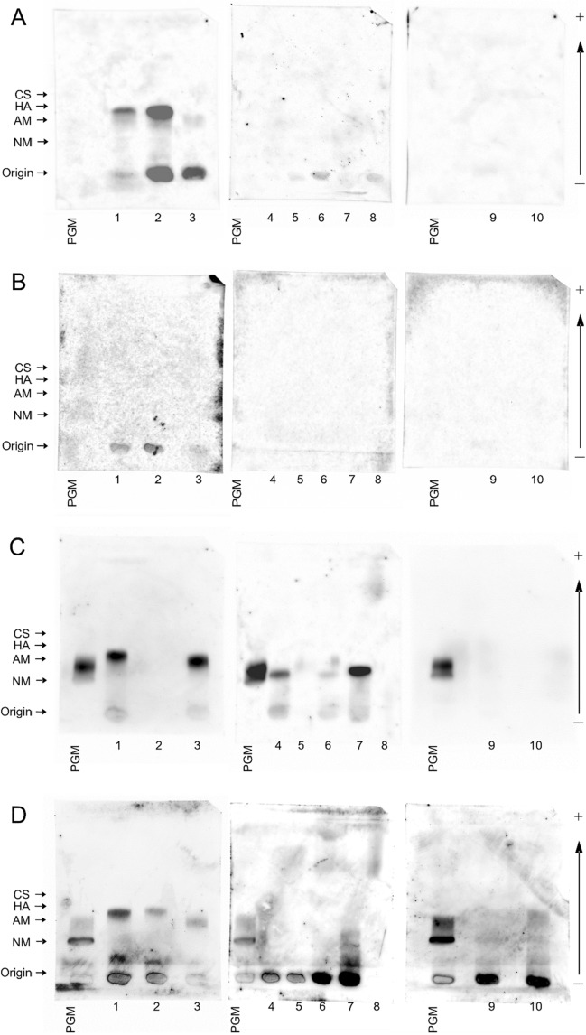
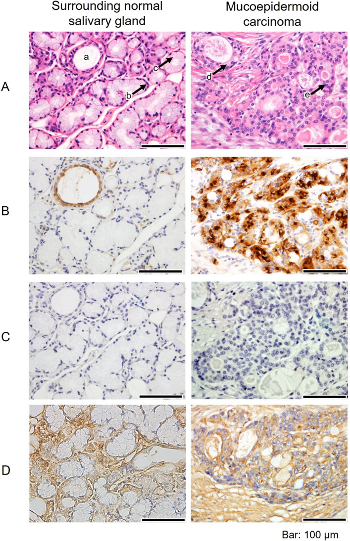
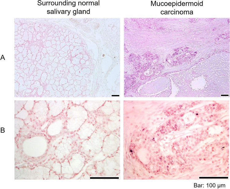
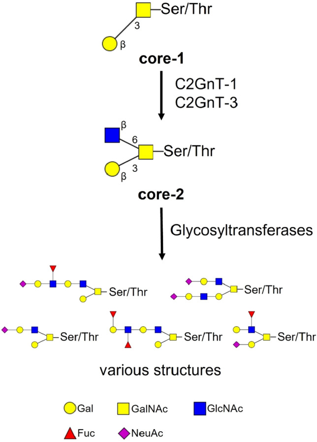
Mucoepidermoid carcinoma (MEC) is the most frequent of the rare salivary gland malignancies. We previously reported high expression of Mucin 1 (MUC1) modified with sialylated core-2 O-glycans in MEC by using tissue homogenates. In this study, we characterised glycan structures of MEC and identified the localisation of cells expressing these distinctive glycans on MUC1. Mucins were extracted from the frozen tissues of three patients with MEC, and normal salivary glands (NSGs) extracted from seven patients, separated by supported molecular matrix electrophoresis (SMME) and the membranes stained with various lectins. In addition, formalin-fixed, paraffin-embedded sections from three patients with MEC were subjected to immunohistochemistry (IHC) with various monoclonal antibodies and analysed for C2GnT-1 expression by in situ hybridisation (ISH). Lectin blotting of the SMME membranes revealed that glycans on MUC1 from MEC samples contained α2,3-linked sialic acid. In IHC, MUC1 was diffusely detected at MEC-affected regions but was specifically detected at apical membranes in NSGs. ISH showed that C2GnT-1 was expressed at the MUC1-positive in MEC-affected regions but not in the NSG. MEC cells produced MUC1 modified with α2,3-linked sialic acid-containing core-2 O-glycans. MUC1 containing these glycans deserves further study as a new potential diagnostic marker of MEC.
© 2023. The Author(s).
Conflict of interest statement
The authors declare no competing interests.
Figures
Similar articles
-
Characterization of tumor-associated MUC1 and its glycans expressed in mucoepidermoid carcinoma.Oncol Lett. 2021 Oct;22(4):702. doi: 10.3892/ol.2021.12963. Epub 2021 Aug 3. Oncol Lett. 2021. PMID: 34457057 Free PMC article.
-
Expression of membrane-bound mucins (MUC1 and MUC4) and secreted mucins (MUC2, MUC5AC, MUC5B, MUC6 and MUC7) in mucoepidermoid carcinomas of salivary glands.Am J Surg Pathol. 2005 Jun;29(6):806-13. doi: 10.1097/01.pas.0000155856.84553.c9. Am J Surg Pathol. 2005. PMID: 15897748
-
MUC1, MUC2, MUC4, and MUC5AC expression in salivary gland mucoepidermoid carcinoma: diagnostic and prognostic implications.Am J Surg Pathol. 2005 Jul;29(7):881-9. doi: 10.1097/01.pas.0000159103.95360.e8. Am J Surg Pathol. 2005. PMID: 15958852
-
Diverse glycosylation of MUC1 and MUC2: potential significance in tumor immunity.J Biochem. 1999 Dec;126(6):975-85. doi: 10.1093/oxfordjournals.jbchem.a022565. J Biochem. 1999. PMID: 10578046 Review.
-
Expression of mucin antigens in human cancers and its relationship with malignancy potential.Pathol Int. 1997 Dec;47(12):813-30. doi: 10.1111/j.1440-1827.1997.tb03713.x. Pathol Int. 1997. PMID: 9503463 Review.
Cited by
-
Preparation of Soluble Mucin Solutions from the Salivary Glands.Methods Mol Biol. 2024;2763:45-50. doi: 10.1007/978-1-0716-3670-1_3. Methods Mol Biol. 2024. PMID: 38347398
References
-
- Suhela R, Gattu V. Study of morphological subtypes of major salivary gland tumors. Perspect. Med. Res. 2017;3:24–28.
-
- Naggar AK, et al. WHO Classification of Head and Neck Tumors. 4. World Health Organization; 2017. pp. 159–164.
Publication types
MeSH terms
Substances
LinkOut - more resources
Full Text Sources
Research Materials
Miscellaneous
