. 2023 Apr 8;6(1):383.
doi: 10.1038/s42003-023-04771-9.

A tailored tetravalent peptide displays dual functions to inhibit amyloid β production and aggregation

Affiliations

A tailored tetravalent peptide displays dual functions to inhibit amyloid β production and aggregation

Waka Sato et al. Commun Biol. .

Abstract

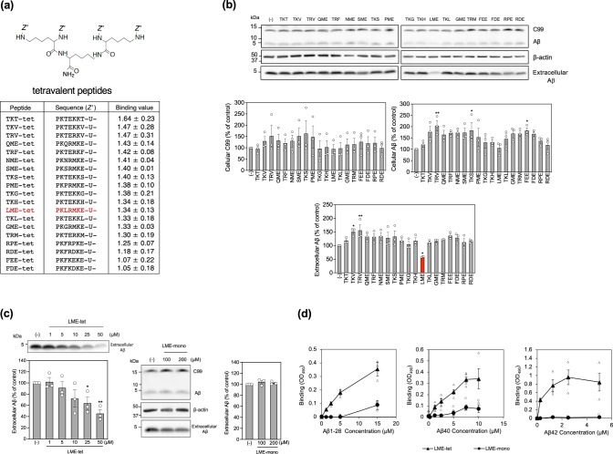
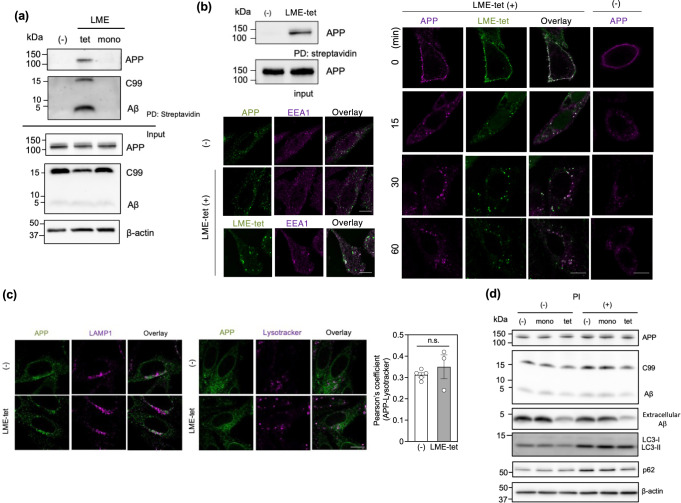
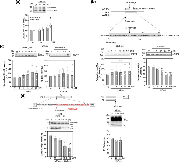
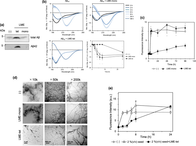
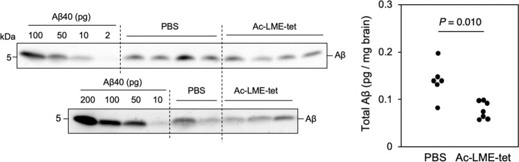
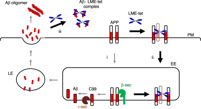
Inhibition of amyloid-β peptide (Aβ) accumulation in the brain is a promising approach for treatment of Alzheimer's disease (AD). Aβ is produced by β-secretase and γ-secretase in endosomes via sequential proteolysis of amyloid precursor protein (APP). Aβ and APP have a common feature to readily cluster to form multimers. Here, using multivalent peptide library screens, we identified a tetravalent peptide, LME-tet, which binds APP and Aβ via multivalent interactions. In cells, LME-tet-bound APP in the plasma membrane is transported to endosomes, blocking Aβ production through specific inhibition of β-cleavage, but not γ-cleavage. LME-tet further suppresses Aβ aggregation by blocking formation of the β-sheet conformation. Inhibitory effects are not observed with a monomeric peptide, emphasizing the significance of multivalent interactions for mediating these activities. Critically, LME-tet efficiently reduces Aβ levels in the brain of AD model mice, suggesting it may hold promise for treatment of AD.


Conflict of interest statement

The authors declare no competing interests.

Figures

Fig. 1
Fig. 1. Affinity-driven screening of tetravalent peptide libraries synthesized on a membrane identified a series of Aβ1–28 binding peptides.
a Schematic diagram of amyloid precursor protein (APP), C-terminal fragment β (C99), and amyloid-β peptide (Aβ). b Schematic showing the structure of our tetravalent peptide library synthesized on a cellulose membrane. The library for the first round of screening contained tetravalent peptides with the sequence X-X-X-X-X-X-X-U- (where X is a mixture of all amino acids except Cys, and U is amino hexanoic acid as a spacer), and each X was replaced individually by a fixed amino acid, excluding Cys (Supplementary Fig. 1a). Membranes were blotted with biotinylated Aβ1–28 (10 µg/ml), and binding of Aβ1–28 to each tetravalent peptide was analyzed, as described in detail in the Methods and in Supplementary Fig. 1a. c Schematic outline of the affinity-based maturation of Aβ1–28-binding motifs. The second library was designed based on the motif obtained from the first screen. Similarly, five additional rounds of affinity-based screening were performed to maturate the binding motifs and generate the seventh libraries (Supplementary Fig. 1b–j). d The library for the seventh round of screening contained peptides with following sequences: P-K-K-X-M-K-E-U- (7th Ic), P-K-X-R-M-K-E-U- (7th Id), P-K-F-K-X-K-E-U- (7th Ie), P-K-F-R-X-K-E-U- (7th If), P-K-T-E-K-K-X-U- (7th IIa), or P-K-T-E-R-K-X-U- (7th IIb), where each X was replaced individually by a fixed amino acid, other than Cys; “ori” indicates the original motif. The binding of Aβ1–28 to each tetravalent peptide was analyzed, as described above, and each screen was performed three times; a representative blotted membrane is shown (upper panel). Mean Aβ-binding values for the top 60 motifs, ± the standard error (SE), are shown in the lower panel (n = 3). The top 15 motifs were selected as candidates, and four additional motifs were selected from groups Ie and If, as no motifs from these groups were represented in the top 15; selected motifs are boxed in red.
Fig. 2
Fig. 2. LME-tet efficiently inhibits production of Aβ in cells through a multivalent interaction.
a Schematic showing the structure of the tetravalent peptides and the Aβ1–28-binding motifs identified by the seventh round of screening. b Effects of tetravalent peptides on production of Aβ. 7WD10 Chinse Hamster Ovary (CHO) cells were treated with each tetravalent peptide (50 µM) for 48 h; cell lysates (for intracellular C99 and Aβ) and culture medium (for extracellular Aβ) from peptide-treated and vehicle control-treated cells were analyzed by western blot. Data are graphed as the percentage of the control value, showing the mean ± SE; n = 3. Significance vs. vehicle was calculated by analysis of variance (ANOVA), followed by one-sided Dunnett’s test; *P < 0.05, **P < 0.01. c Effect of increasing concentrations of LME-tet on Aβ production (left panel). 7WD10 cells were treated with vehicle control or LME-tet at the indicated concentrations for 48 h, and the culture medium was analyzed by western blot. Data are graphed as the percentage of the control value, showing the mean ± SE; n = 3. Significance vs. vehicle was calculated by ANOVA, followed by one-sided Dunnett’s test; *P < 0.05, **P < 0.01. Effect of LME-mono on Aβ production (right panels). 7WD10 cells were treated with vehicle control or LME-mono at the indicated concentrations for 48 h; cell lysates (for intracellular C99 and Aβ) and culture medium (for extracellular Aβ) were analyzed by western blot. LME-mono concentrations of 100 and 200 µM contain the same moles of binding motif as LME-tet at 25 and 50 µM, respectively. Data are graphed as the percentage of the control value, showing the mean ± SE; n = 3. Significance vs. vehicle was calculated by ANOVA, followed by one-sided Dunnett’s test; *P < 0.05. d Binding of LME-tet and LME-mono to Aβ1-28, Aβ40 and Aβ42 was measured by enzyme-linked immunosorbent assay (ELISA). Values represent the mean ± SE; n = 3 for Aβ1–28 and Aβ42, n = 4 for Aβ40.
Fig. 3
Fig. 3. LME-tet forms a complex with APP and co-localizes with APP in endosomes.
a Binding of LME-tet to intracellular APP, C99, and Aβ. 7WD10 cells were treated with biotinylated LME-tet (50 µM) or LME-mono (200 µM) for 30 min at 37 °C. Cell lysates were then incubated with streptavidin beads, and coprecipitating proteins were analyzed by western blot; PD, pull down. b Binding of LME-tet to APP in the plasma membrane (upper left panel). 7WD10 cells were treated with biotinylated LME-tet (50 µM) on ice for 30 min, and cell lysates were analyzed as described above. Intracellular localization of APP-LME-tet complex (right panel). 7WD10 cells were treated with mouse monoclonal anti-human APP antibody 6E10 in the presence or absence of biotinylated LME-tet (50 µM) on ice for 30 min; cells were then washed and incubated at 37 °C for the indicated time. APP was detected by Alexa Fluor 546-conjugated goat anti-mouse IgG antibody, and LME-tet was detected by Alexa Fluor 488-conjugated streptavidin. Colocalization with early endosome antigen 1 (EEA1; lower left panel). 7WD10 cells were treated as described above, and after 15 min incubation at 37 °C, immunostaining for EEA1 was performed using rabbit polyclonal anti-EEA1 antibody, followed by Alexa Fluor 488-conjugated goat anti-rabbit IgG antibody. The scale bars indicate 10 µm. c APP co-localization with lysosome-associated membrane glycoprotein 1 (LAMP1), a lysosomal marker, was analyzed by immunocytochemical staining with rabbit polyclonal anti-LAMP1 antibody, followed by Alexa Fluor 488-conjugated goat anti-rabbit IgG antibody (left panel). Acidification of APP-containing endosomes was analyzed using Lysotracker (middle panel). In both cases, 7WD10 cells were treated with or without LME-tet (25 µM) for 60 min at 37 °C, and APP was detected as described above. The scale bars indicate 10 µm. Pearson’s coefficient for colocalization between APP and Lysotracker is shown (right panel; mean ± SE, n (number of cells) = 10–24 or 7–14, for the absence (-) from five independent experiments or presence of LME-tet from three independent experiments, respectively). Pearson’s coefficient was calculated using the Coloc 2 plugin in Image J. The significance vs. LME-tet (-) was calculated using ANOVA, followed by a one-sided Dunnett’s test. n.s. not significant. d Effect of protease inhibitor (PI) on levels of APP, C99, Aβ, and the autophagosomal markers LC3 and p62, in LME-tet- and LME-mono-treated cells. 7WD10 cells were treated with LME-tet (25 µM) or LME-mono (100 µM) in the presence or absence of lysosomal PI cocktail for 48 h. Cell lysates (for intracellular APP, C99, Aβ, LC3, and p62) and culture medium (for extracellular Aβ) were analyzed by western blot.
Fig. 4
Fig. 4. LME-tet specifically inhibits β-secretase to block production of Aβ.
a Effect of LME-tet on APP production. 7WD10 cells were treated with vehicle control or LME-tet at the indicated concentrations for 48 h. Cell lysates were analyzed by western blot. b Effect of LME-tet on production of extracellular sAPPα and sAPPβ Schematic showing α- and β-cleavage sites (top). 7WD10 cells were treated as in (a) and sAPPα and sAPPβ in culture medium were analyzed by western blot (bottom left and right panels, respectively). a, b APP and sAPPα were detected using anti-APP antibody 6E10, and sAPPβ was detected using anti-sAPPβ antibody. Data are graphed as the percentage of the control value, showing the mean ± SE; n = 3–4. Significance vs. vehicle was calculated by ANOVA, followed by one-sided Dunnett’s test; *P < 0.05, **P < 0.01, ***P < 0.001. c Effect of LME-tet on β-cleavage of β-galactoside α-2,6- sialyltransferase 1 (St6gal1) and γ-cleavage of NotchΔE. 7WD10-NotchΔE-St6gal1 cells were treated with LME-tet at the indicated concentrations and β-secretase inhibitor II (βi; 10 µM) for 48 h; culture medium was analyzed by western blot using anti-St6gal1 antibody (left panel). 7WD10-NotchΔE cells were treated with LME-tet at the indicated concentrations and γ-secretase inhibitor DAPT (γi; 10 µM) for 48 h; cell lysates were analyzed by western blot using anti-Myc antibody (right panel). Data are graphed as the percentage of the control value, showing the mean ± SE; n = 3–4. Significance vs. vehicle was calculated by ANOVA, followed by one-sided Dunnett’s test. d Effect of LME-tet on β- and γ-secretase activities in vitro. Schematics showing β- and γ-cleavage sites/products and FLAG-tag locations (top left and right panels, respectively). Purified FLAG-tagged APP fragment (APP633–685-FLAG) with N-terminal Myc and C-terminal FLAG tags was incubated with β-secretase and LME-tet/LME-mono at the indicated concentrations for 4 h (left). FLAG-tagged C99 was incubated with γ-secretase and LME-tet at the indicated concentrations (right). Aβ33-FLAG (left) and Aβ (right) were measured by western blot using anti-Aβ antibody 82E1. APP633–685-FLAG (left) was measured by western blot using anti-Aβ antibody 6E10. Graphs show the mean ± SE, n = 3. Significance vs. vehicle was calculated by ANOVA, followed by one-sided Dunnett’s test; **P < 0.01, ***P < 0.001.
Fig. 5
Fig. 5. LME-tet inhibits the conformational change and subsequent fibrillation of Aβ42.
a Binding of LME-tet to extracellular Aβ. Culture medium from 7WD10 cells recovered after 48 h of growth was treated with biotinylated LME-tet (50 µM) or LME-mono (200 µM). Medium was then incubated with streptavidin beads, and coprecipitating Aβ40 and Aβ42 were analyzed by western blot using anti-Aβ or anti-Aβ42-specific antibody. b Time-dependent conformational change in Aβ42 (20 µM) in the presence of LME-tet (2 µM, lower left panel) or LME-mono (2 µM, upper right panel). Far-UV circular dichroism (CD) spectra (wavelength 190–250 nm) were measured at the indicated time points. Kinetic rates of β-sheet formation of Aβ42 were estimated by measuring time-dependent alterations of molar ellipticities of CD spectra at 217 nm (lower right panel). Data represent the mean ± SE; n = 3–4. c Fibrillation profiles of Aβ42 in the presence of LME-tet (2 µM) or LME-mono (2 µM), measured by Thioflavin T-binding assay. Time-dependent increments of Aβ42–Thioflavin T binding are shown. Data represent the mean ± SE; n = 3. d Fibrillation profiles of Aβ42 in the presence of LME-tet (2 µM) or LME-mono (2 µM), analyzed by electron microscopy; representative images at ×10 K, ×50 K, and ×200 K magnification (left, right, and center, respectively) are shown. Scale bars indicate 2 µm, 400 nm, and 100 nm, respectively. e Effects of LME-tet (2 µM) on growth of Aβ42 fibril in the presence of 2% (v/v) Aβ42 fibril-seed, measured by the Thioflavin T-binding assay. Time-dependent increments of Aβ42–Thioflavin T binding are shown. Data represent the mean ± SE; n = 3.
Fig. 6
Fig. 6. Ac-LME-tet reduces Aβ levels in the brain of AD model mice.
AppNL-G-F/NL-G-F mice were intraperitoneally administered N-terminal-acetylated LME-tet (Ac-LME-tet) or phosphate-buffered saline (PBS) control. After 7 days, mice were then sacrificed, and the amounts of human Aβ present in the cerebral cortex and the olfactory bulb were measured by western blot using anti-human Aβ antibody 4G8. The indicated amounts of Aβ were used for quantification; n = 6 for control, and n = 7 for Ac-LME-tet-treated mice. Significance was determined by the Mann–Whitney U test; P = 0.010 for Ac-LME-tet-treated vs. PBS-treated mice.
Fig. 7
Fig. 7. Model for the proposed molecular mechanism underlying the intracellular and extracellular effects of LME-tet.
APP present in the plasma membrane (PM) is internalized into early endosomes (EE) and then into late/recycling endosomes (LE), where Aβ is produced by sequential proteolysis of APP performed by β-secretase and γ-secretase. Aβ is released into the extracellular space and then aggregates to from Aβ oligomers (i, gray arrows). LME-tet binds to APP in the plasma membrane through multivalent interaction. This complex is transported to the endosomes and blocks Aβ production through specific inhibition of β-cleavage, but not γ-cleavage (ii). In addition, LME-tet suppresses oligomerization of extracellular Aβ by inhibiting the formation of its β-sheet conformation (iii).

Similar articles

Cited by

References

    1. Masters CL, et al. Amyloid plaque core protein in Alzheimer disease and Down syndrome. Proc. Natl. Acad. Sci. USA. 1985;82:4245–4249. doi: 10.1073/pnas.82.12.4245. - DOI - PMC - PubMed
    1. Long JM, Holtzman DM. Alzheimer disease: an update on pathobiology and treatment strategies. Cell. 2019;179:312–339. doi: 10.1016/j.cell.2019.09.001. - DOI - PMC - PubMed
    1. Kang J, et al. The precursor of Alzheimer’s disease amyloid A4 protein resembles a cell-surface receptor. Nature. 1987;325:733–736. doi: 10.1038/325733a0. - DOI - PubMed
    1. Haass C, Kaether C, Thinakaran G, Sisodia S. Trafficking and proteolytic processing of APP. Cold Spring Harb. Perspect. Med. 2012;2:a006270. doi: 10.1101/cshperspect.a006270. - DOI - PMC - PubMed
    1. Selkoe DJ, et al. The role of APP processing and trafficking pathways in the formation of amyloid beta-protein. Ann. N. Y. Acad. Sci. 1996;777:57–64. doi: 10.1111/j.1749-6632.1996.tb34401.x. - DOI - PubMed

Publication types

MeSH terms

Substances