. 2023 Apr 8;6(1):385.
doi: 10.1038/s42003-023-04763-9.

The role of RNF149 in the pre-emptive quality control substrate ubiquitination

Affiliations

The role of RNF149 in the pre-emptive quality control substrate ubiquitination

Aster Legesse et al. Commun Biol. .

Abstract

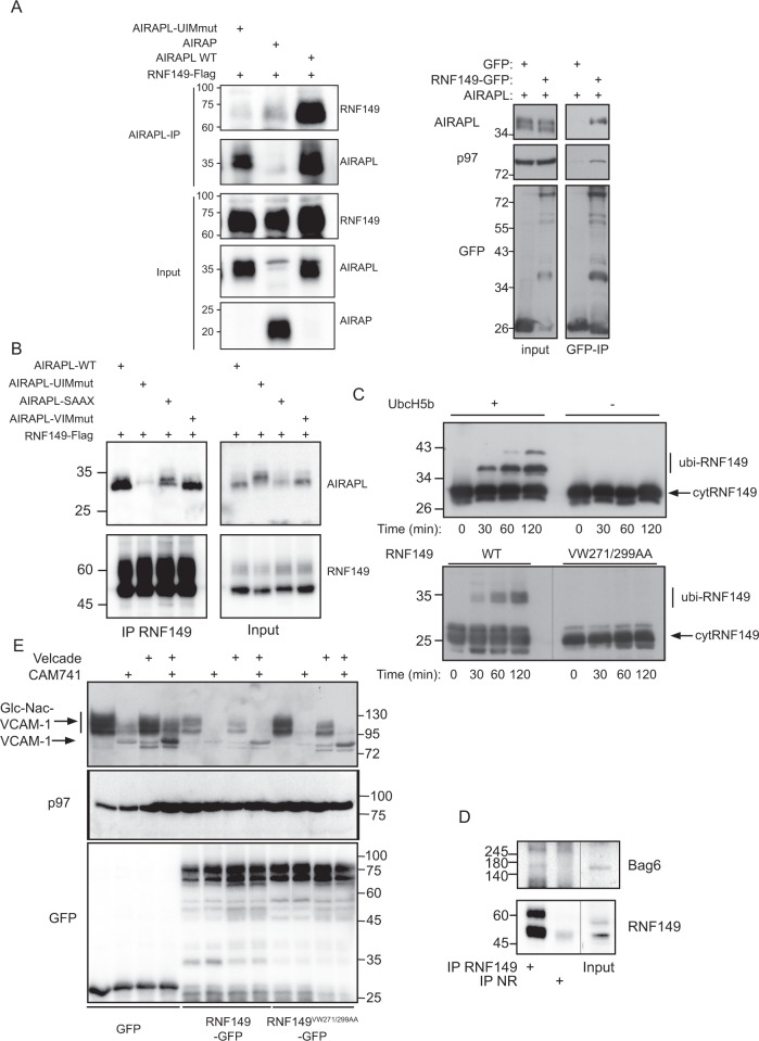
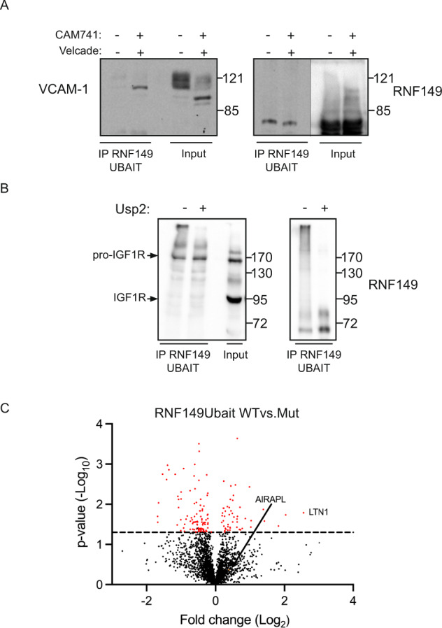
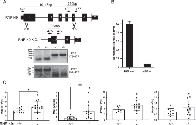
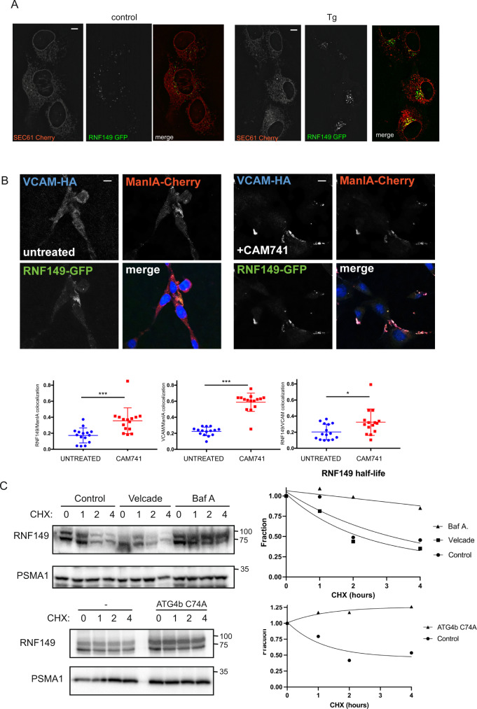
Protein quality control is a process in which a protein's folding status is constantly monitored. Mislocalized proteins (MLP), are processed by the various quality control pathways, as they are often misfolded due to inappropriate cellular surroundings. Polypeptides that fail to translocate into the ER due to an inefficient signal peptide, mutations or ER stress are recognized by the pre-emptive ER associated quality control (pEQC) pathway and degraded by the 26 S proteasome. In this report we reveal the role of RNF149, a membrane bound E3 ligase in the ubiquitination of known pEQC substrates. We demonstrate its selective binding only to non-translocated proteins and its association with known pEQC components. Impairment in RNF149 function increases translocation flux into the ER and manifests in a myeloproliferative neoplasm (MPN) phenotype, a pathological condition associated with pEQC impairment. Finally, the dynamic localization of RNF149 may provide a molecular switch to regulate pEQC during ER stress.


Conflict of interest statement

The authors declare no competing interests.

Figures

Fig. 1
Fig. 1. RNF149 binding to pEQC components.
A Left-293 cell lysates expressing RNF149-Flag alongside the indicated AIRAPL isoform or AIRAP (used as a specificity control), were immunopurified (IP) with AIRAP or AIRAPL antibodies and RNF149 content was revealed with a Flag immunoblot. Total content prior to IP is indicated as input. Right- 293 cell lysates expressing AIRAPL alongside the indicated RNF149-GFP or GFP (used as a specificity control), were IPed with a GFP antibody. AIRAPL and endogenous p97 content were revealed by immunoblot. Total content prior to IP is indicated as input. B 293 cell lysates expressing RNF149 alongside the indicated AIRAPL isoforms were IPed with an RNF149 antibody. AIRAPL content was revealed by immunoblot. Total content prior to IP is indicated as input. C A time course in-vitro ubiquitin ligase assay using recombinant cytosolic portion of RNF149 (AA222-400) as a E3 ligase, in the presence or absence of the E2 UbcH5b (upper panel) or a RING mutant VW271/299AA (lower panel). D Binding of endogenous RNF149 to endogenous Bag6 was evaluated by an IP of RNF149 or a non-relevant (N.R.) antibody IP from 293 cells stably expressing the pEQC substrate VCAM1-YFP. Bag6 content was revealed by immunoblot. Total content prior to IP is indicated as input. E Cells expressing VCAM-1 alongside GFP, RNF149WT-GFP or RNF149RINGmut-GFP were treated with CAM741 or velcade as indicated. The presence of the non-translocated (VCAM-1) and glycosylated translocated protein (Glc-NAC-VCAM-1) are indicated. GFP immunoblot show the expression level of the free or fused RNF149 forms and p97 IB serves as a loading control of lysates.
Fig. 2
Fig. 2. pEQC substrate association with RNF149.
A 293 cells expressing RNF149UBAIT and the pEQC substrate VCAM-1 were treated as indicated with velcade and CAM741. Cell lysates were adjusted to RIPA buffer and a Flag IP was applied followed by extensive RIPA buffer washes. IP content was evaluated towards RNF149 and VCAM-1 content by Flag and HA immunoblots respectively. Total content prior to IP is indicated as input. B 293 cells expressing IGF1R-GFP and RNF149UBAIT were subjected to a Flag IP under RIPA conditions. Following extensive RIPA buffer washes the IP content was split into two equal fractions and the deubiquitinating enzyme Usp2 was added to half of the purified content. Deubiquitintion was terminated by addition of laemmli buffer and RNF149 and IGF1R content was evaluated by Flag (RNF149) and GFP (IGF-1R) immunoblots. C 293 cells expressing RNF149UBAIT wt or mutant forms were purified from cell extracts and protein content identification was evaluated by LC-MS. A volcano plot showing the enrichment fold (X-axis) and significance (Y-axis) is shown. In addition to AIRAPL, the top ranked specific interactor with RNF149UBAIT wt is labeled (LTN1).
Fig. 3
Fig. 3. RNF149 knockout phenotype.
A Mouse genomic locus illustration of RNF149. Forward and reverse primer binding sites (475, 492 and 477) are shown as well as scissor icons indicating the designed Cas9 cleavage sites. Expected PCR product size of the primer sets are indicated. Genotyping examples of RNF149 + / + and RNF149-/- animals using the designated primer sets shown on bottom. PCR products were sequenced and confirmed the deletion of the 19 kb RNF149 genomic fragment. B RT-PCR results of cDNA samples from the indicated genotypes were performed on MEF cells from the respective mice. RNA levels were normalized to a housekeeping gene (HPRT1). C Blood cells extracted from the indicated RNF149 genotype were evaluated at the age of 6 month. WBC-white blood cells; Neu-neutrophils; Mon-monocyte; Lym-lymphocyte. *p < 0.05; **p < 0.01.
Fig. 4
Fig. 4. RNF149 knockout impact on translocation.
A Left- Illustration of VCAM-1 containing the Flag signal sequence (FSS), the glycosylation sites, transmembrane domain (TM), and C-terminus HA tag. Right-Cells expressing Flag-VCAM1-HA were untreated or subjected to velcade and CAM741 treatment. The HA immunoblot reveals both ER and cytosol species of VCAM1 whereas the Flag immunoblot reveals only the pEQC non-translocated species that harbors the signal sequence. The lower band that is not recognized by the Flag immunoblot is an ER translocated species whose signal sequence has been cleaved. Proteasomal stabilization of this species (also seen in Fig. 1C with only velcade) indicates that a small percentage of VCAM1 is also subjected to ERAD. B RNF149 wildtype and knockout MEF cells stably expressing VCAM1 were treated with CAM741 and velcade as indicated. Steady state levels of VCAM1 were revealed by a HA immunoblot and ratios of pEQC or ERAD were quantified and standard deviations from three repetitions are presented.
Fig. 5
Fig. 5. RNF149 regulation as means of tuning quality control.
A Live cell imaging of cells expressing RNF149-GFP and ER translocon marker Sec61beta-Cherry under control and ER stress conditions. The partial co-localization of RNF149 with Sec61 seems to be induced during ER stress as apparent from the enhanced co-localization (yellow) observed under thapsigargin (Tg) conditions (120 min, 400 nM). Representative images are presented, scale bars are 10um. B Immunofluorescence of cells expressing RNF149-GFP, VCAM-HA and ManIA-Cherry show a disperse pattern of VCAM and a partial co-localization of RNF149 and ManIA. Treatment with CAM741 caused accumulation of VCAM at the QCVs, together with ManIA and RNF149. Nuclear staining was performed by DAPI staining and added to the merged figure. Scale bars are 10um. C Half-life evaluations of RNF149 were evaluated by treating 293 cells expressing RNF149 with CHX for the indicated time points. The evaluation was performed in the presence or absence of velcade or bafilomycin A (top) or in the presence or absence of a catalytic inactive form of ATG4b (bottom). A quantification of the RNF149 fraction remaining along the four hour time course, is indicated (right).

References

    1. Kadowaki H, et al. Pre-emptive quality control protects the ER from protein overload via the proximity of ERAD components and SRP. Cell Rep. 2015;13:944–956. doi: 10.1016/j.celrep.2015.09.047. - DOI - PubMed
    1. Kang SW, et al. Substrate-specific translocational attenuation during ER stress defines a pre-emptive quality control pathway. Cell. 2006;127:999–1013. doi: 10.1016/j.cell.2006.10.032. - DOI - PMC - PubMed
    1. Rane NS, Kang SW, Chakrabarti O, Feigenbaum L, Hegde RS. Reduced translocation of nascent prion protein during ER stress contributes to neurodegeneration. Dev. Cell. 2008;15:359–370. doi: 10.1016/j.devcel.2008.06.015. - DOI - PMC - PubMed
    1. Cox JS, Walter P. A novel mechanism for regulating activity of a transcription factor that controls the unfolded protein response. Cell. 1996;87:391–404. doi: 10.1016/S0092-8674(00)81360-4. - DOI - PubMed
    1. Haze K, Yoshida H, Yanagi H, Yura T, Mori K. Mammalian transcription factor ATF6 is synthesized as a transmembrane protein and activated by proteolysis in response to endoplasmic reticulum stress. Mol. Biol. Cell. 1999;10:3787–3799. doi: 10.1091/mbc.10.11.3787. - DOI - PMC - PubMed

Publication types

MeSH terms

Substances

LinkOut - more resources