

Therapeutic effects of anti-GM2 CAR-T cells expressing IL-7 and CCL19 for GM2-positive solid cancer in xenograft model
- PMID: 37031457
- PMCID: PMC10278466
- DOI: 10.1002/cam4.5907
Therapeutic effects of anti-GM2 CAR-T cells expressing IL-7 and CCL19 for GM2-positive solid cancer in xenograft model
Abstract
Background: While chimeric antigen receptor (CAR)-T cell therapy has demonstrated excellent efficacy in hematopoietic malignancies, its clinical application in solid cancers has yet to be achieved. One of the reasons for such hurdle is a lack of suitable CAR targets in solid cancers.
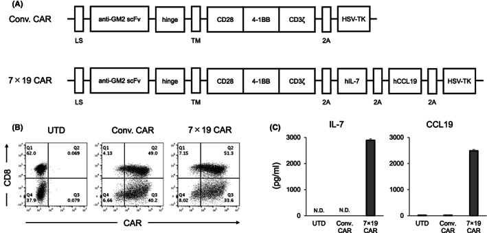
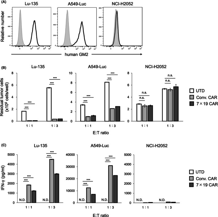
Methods: GM2 is one of the gangliosides, a group of glycosphingolipids with sialic acid in the glycan, and overexpressed in various types of solid cancers. In this study, by using interleukin (IL)-7 and chemokine (C-C motif) ligand 19 (CCL19)-producing human CAR-T system which we previously developed, a possibility of GM2 as a solid tumor target for CAR-T cell therapy was explored in a mouse model with human small-cell lung cancer.
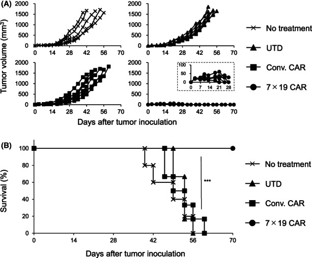
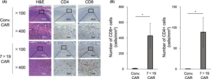
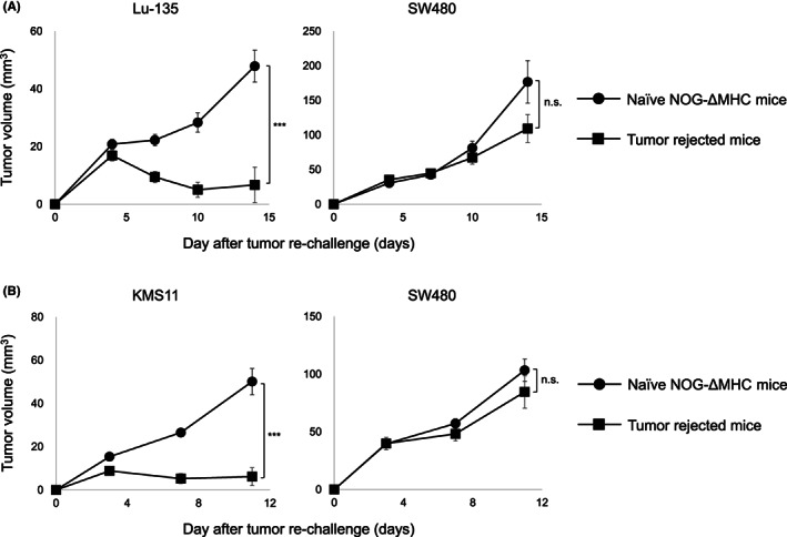
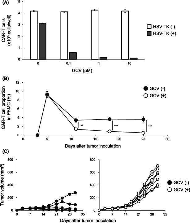
Results: Treatment with anti-GM2 IL-7/CCL19-producing CAR-T cells induced complete tumor regression along with an abundant T cell infiltration into the solid tumor tissue and long-term memory responses, without any detectable adverse events. In addition, as measures to control cytokine-release syndrome and neurotoxicity which could occur in association with clinical use of CAR-T cells, we incorporated Herpes simplex virus-thymidine kinase (HSV-TK), a suicide system to trigger apoptosis by administration of ganciclovir (GCV). HSV-TK-expressing anti-GM2 IL-7/CCL19-producing human CAR-T cells were efficiently eliminated by GCV administration in vivo.
Conclusions: Our study revealed the promising therapeutic efficacy of anti-GM2 IL-7/CCL19-producing human CAR-T cells with an enhanced safety for clinical application in the treatment of patients with GM2-positive solid cancers.
Keywords: CAR-T cell; chemokine; cytokine; ganglioside; solid cancers.
© 2023 The Authors. Cancer Medicine published by John Wiley & Sons Ltd.
Conflict of interest statement
Koji Tamada and Yukimi Sakoda hold stocks of Noile‐Immune Biotech and receive remuneration from Noile‐Immune Biotech. Koji Tamada received lecture fees from Ono Pharmaceutical, MSD, and Chugai Pharmaceutical. Koji Tamada received a research fund from Chugai Pharmaceutical. Yukimi Sakoda received a research fund from Noile‐Immune Biotech. Other authors declare no conflict of interest.
Figures
Similar articles
-
Therapeutic Efficacy of IL7/CCL19-Expressing CAR-T Cells in Intractable Solid Tumor Models of Glioblastoma and Pancreatic Cancer.Cancer Res Commun. 2024 Sep 1;4(9):2514-2524. doi: 10.1158/2767-9764.CRC-24-0226. Cancer Res Commun. 2024. PMID: 39240078 Free PMC article.
-
Enhanced anti-tumor efficacy of IL-7/CCL19-producing human CAR-T cells in orthotopic and patient-derived xenograft tumor models.Cancer Immunol Immunother. 2021 Sep;70(9):2503-2515. doi: 10.1007/s00262-021-02853-3. Epub 2021 Feb 8. Cancer Immunol Immunother. 2021. PMID: 33559069 Free PMC article.
-
[Construction and function of Glypican-3-targeted fourth-generation chimeric antigen receptor T cells (secreting IL-7 and CCL19)].Sheng Wu Gong Cheng Xue Bao. 2020 May 25;36(5):979-991. doi: 10.13345/j.cjb.200106. Sheng Wu Gong Cheng Xue Bao. 2020. PMID: 32567281 Chinese.
-
Chimeric antigen-receptor T-cell therapy for hematological malignancies and solid tumors: Clinical data to date, current limitations and perspectives.Curr Res Transl Med. 2017 Sep;65(3):93-102. doi: 10.1016/j.retram.2017.08.003. Curr Res Transl Med. 2017. PMID: 28988742 Review.
-
Chimeric antigen receptor-engineered T-cell therapy for liver cancer.Hepatobiliary Pancreat Dis Int. 2018 Aug;17(4):301-309. doi: 10.1016/j.hbpd.2018.05.005. Epub 2018 May 24. Hepatobiliary Pancreat Dis Int. 2018. PMID: 29861325 Review.
Cited by
-
Advances in adoptive cell therapies in small cell lung cancer.Explor Target Antitumor Ther. 2025 Mar 26;6:1002302. doi: 10.37349/etat.2025.1002302. eCollection 2025. Explor Target Antitumor Ther. 2025. PMID: 40160238 Free PMC article. Review.
-
Chimeric Antigen Receptor (CAR) T-Cell Therapy for Patients with Lung Cancer: Current Perspectives.Onco Targets Ther. 2023 Jul 3;16:515-532. doi: 10.2147/OTT.S341179. eCollection 2023. Onco Targets Ther. 2023. PMID: 37425981 Free PMC article. Review.
-
Therapeutic Efficacy of IL7/CCL19-Expressing CAR-T Cells in Intractable Solid Tumor Models of Glioblastoma and Pancreatic Cancer.Cancer Res Commun. 2024 Sep 1;4(9):2514-2524. doi: 10.1158/2767-9764.CRC-24-0226. Cancer Res Commun. 2024. PMID: 39240078 Free PMC article.
-
Enhancing the safety of CAR-T cell therapy: Synthetic genetic switch for spatiotemporal control.Sci Adv. 2024 Feb 23;10(8):eadj6251. doi: 10.1126/sciadv.adj6251. Epub 2024 Feb 23. Sci Adv. 2024. PMID: 38394207 Free PMC article. Review.
-
Current Advances and Challenges in CAR-T Therapy for Hematological and Solid Tumors.Immunotargets Ther. 2025 Jun 27;14:655-680. doi: 10.2147/ITT.S519616. eCollection 2025. Immunotargets Ther. 2025. PMID: 40599347 Free PMC article. Review.
References
-
- Schuster SJ, Bishop MR, Tam CS, et al. Tisagenlecleucel in adult relapsed or refractory diffuse large B‐cell lymphoma. N Engl J Med. 2019;380:45‐56. - PubMed
Publication types
MeSH terms
Substances
LinkOut - more resources
Full Text Sources
Medical
