. 2023 Jul:247:103085.
doi: 10.1016/j.autneu.2023.103085. Epub 2023 Apr 6.

Gamma secretase activity modulates BMP-7-induced dendritic growth in primary rat sympathetic neurons

Affiliations

Gamma secretase activity modulates BMP-7-induced dendritic growth in primary rat sympathetic neurons

Krystal Karunungan et al. Auton Neurosci. 2023 Jul.

Abstract

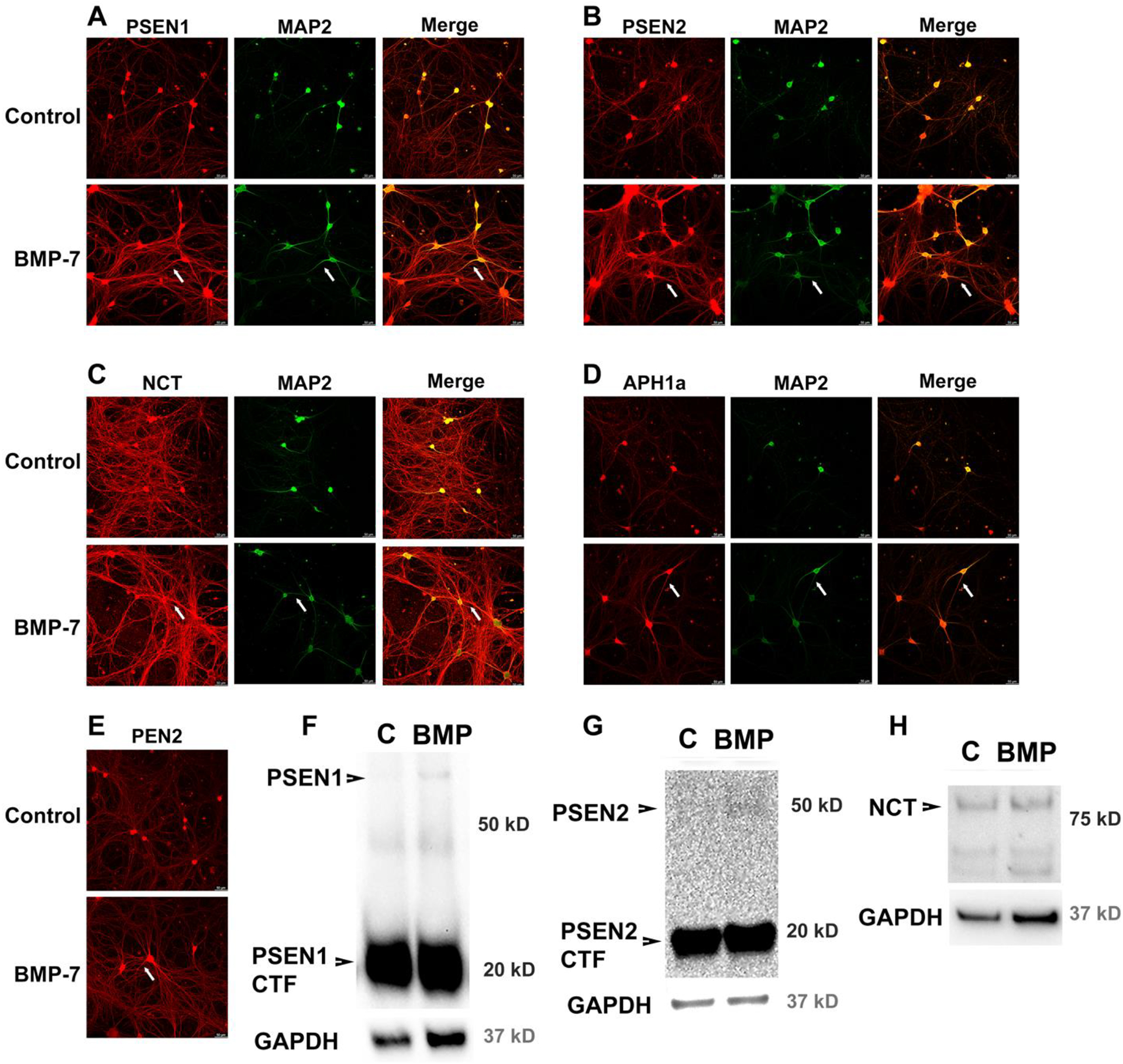
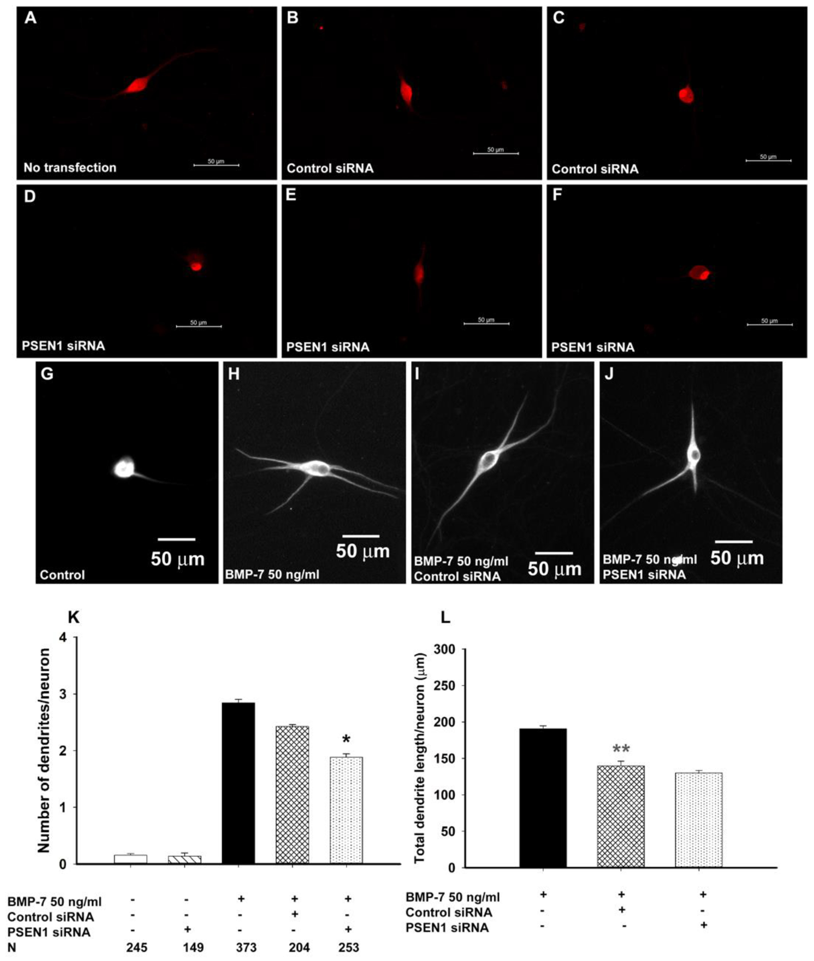
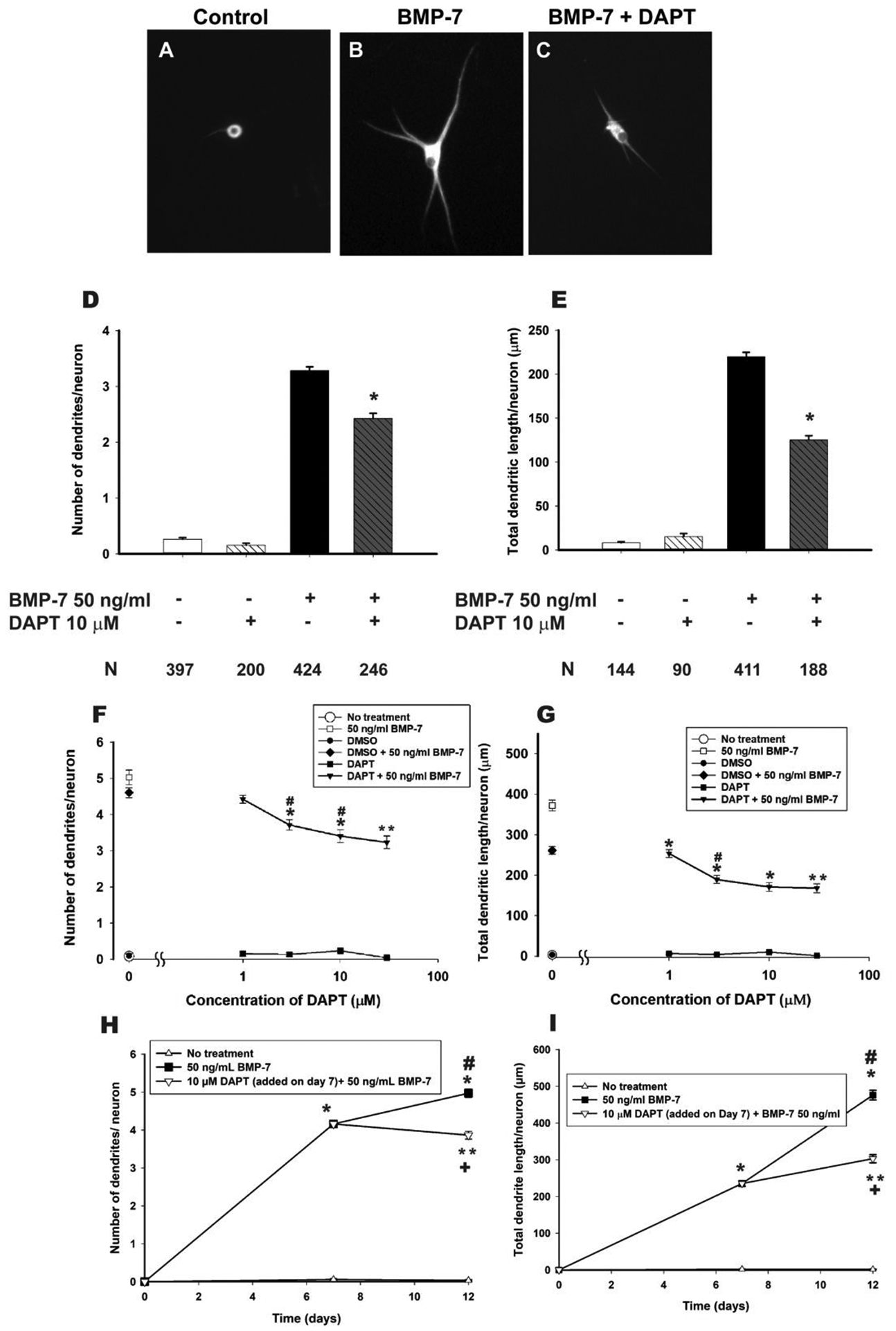
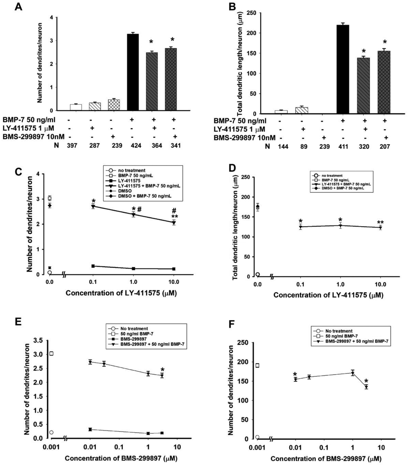
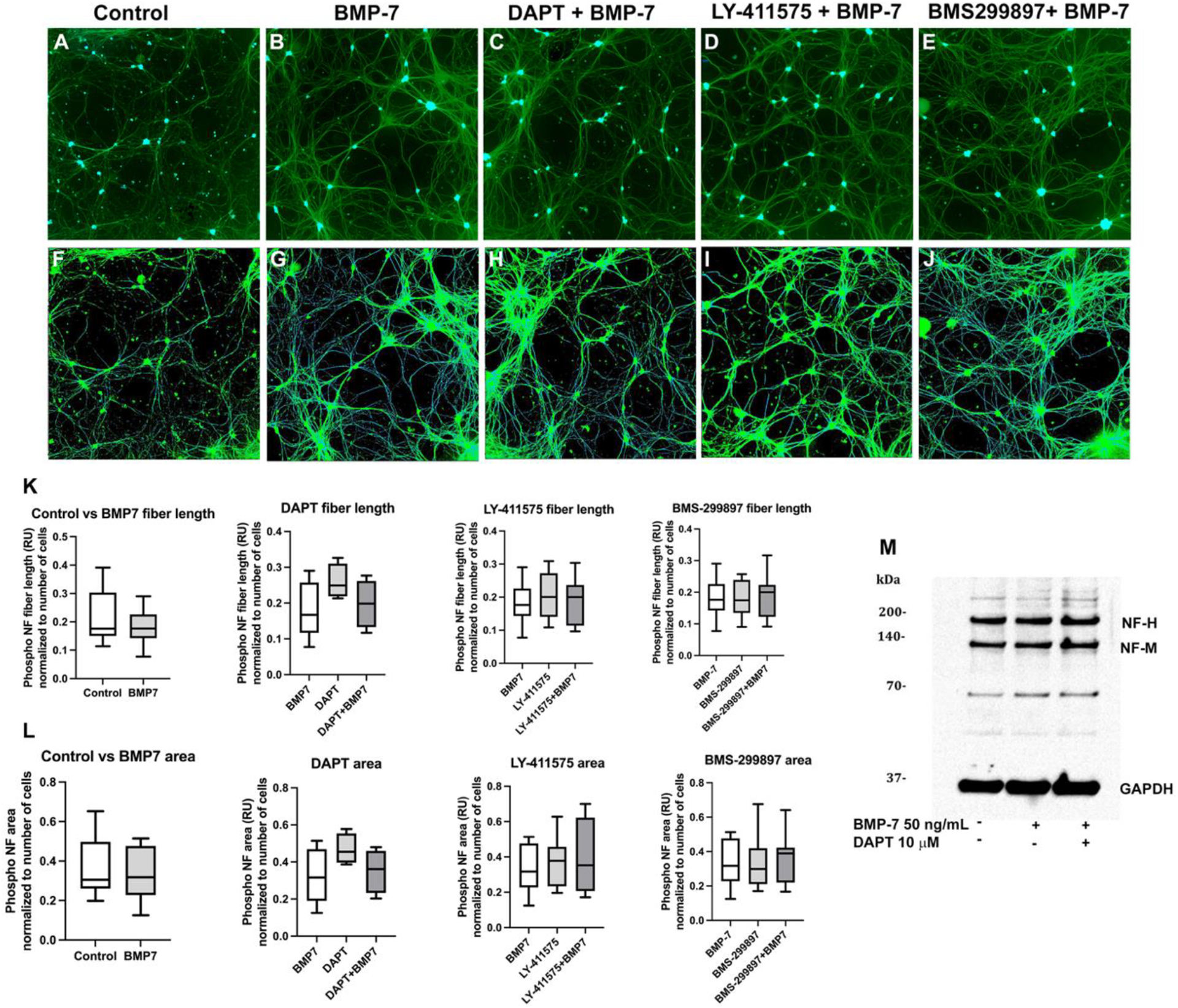
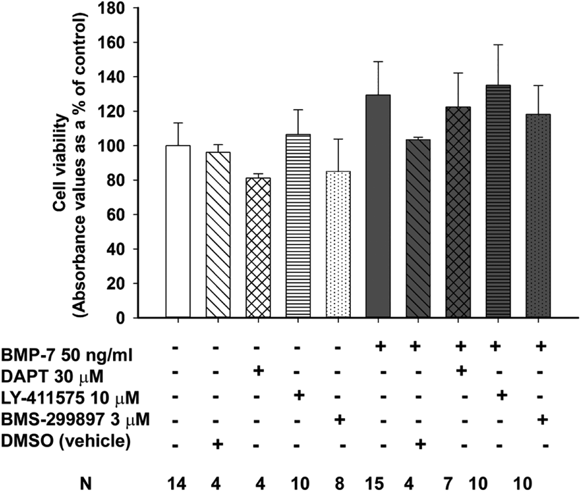
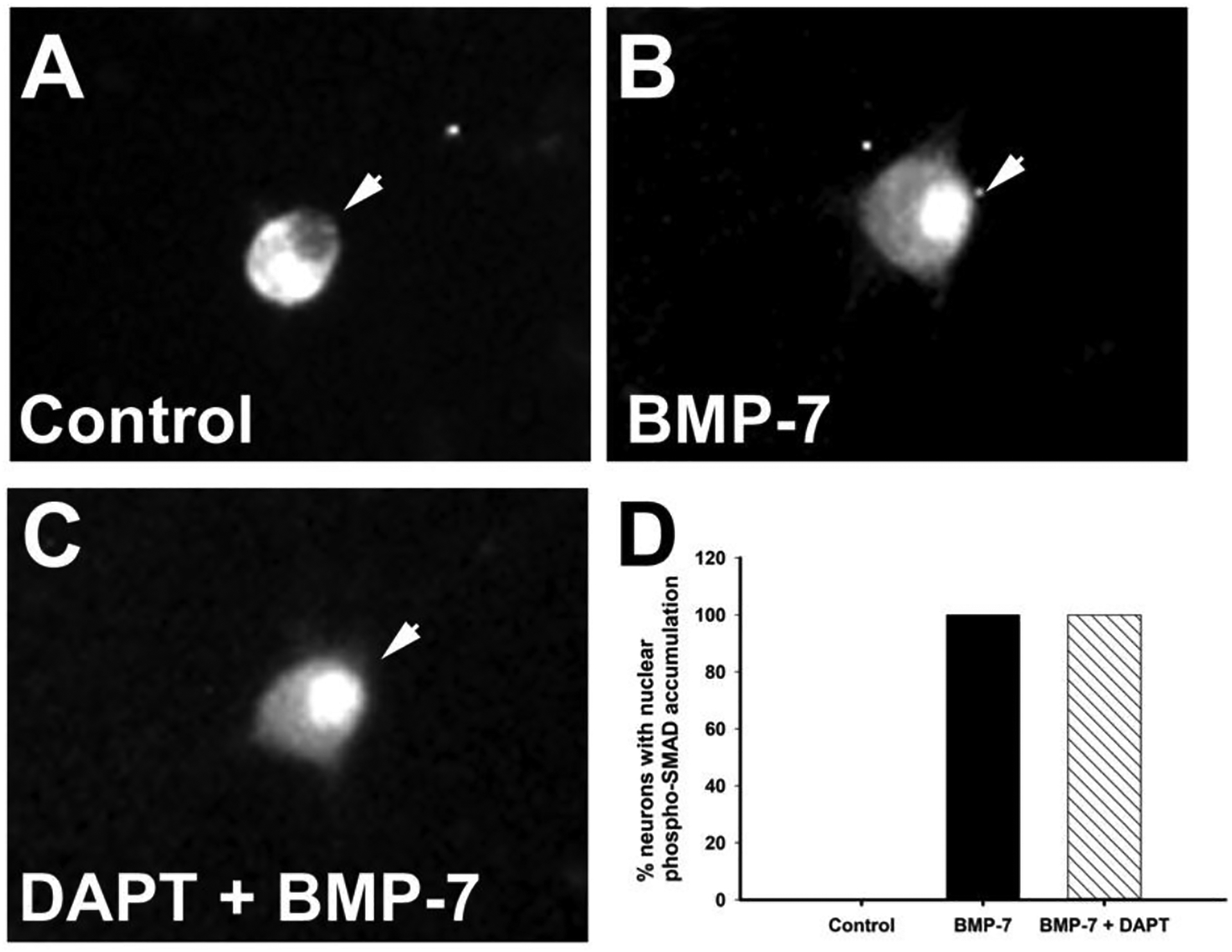
Autonomic dysfunction has been observed in Alzheimer's disease (AD); however, the effects of genes involved in AD on the peripheral nervous system are not well understood. Previous studies have shown that presenilin-1 (PSEN1), the catalytic subunit of the gamma secretase (γ-secretase) complex, mutations in which are associated with familial AD function, regulates dendritic growth in hippocampal neurons. In this study, we examined whether the γ-secretase pathway also influences dendritic growth in primary sympathetic neurons. Using immunoblotting and immunocytochemistry, molecules of the γ-secretase complex, PSEN1, PSEN2, PEN2, nicastrin and APH1a, were detected in sympathetic neurons dissociated from embryonic (E20/21) rat sympathetic ganglia. Addition of bone morphogenetic protein-7 (BMP-7), which induces dendrites in these neurons, did not alter expression or localization of γ-secretase complex proteins. BMP-7-induced dendritic growth was inhibited by siRNA knockdown of PSEN1 and by three γ-secretase inhibitors, γ-secretase inhibitor IX (DAPT), LY-411575 and BMS-299897. These effects were specific to dendrites and concentration-dependent and did not alter early downstream pathways of BMP signaling. In summary, our results indicate that γ-secretase activity enhances BMP-7 induced dendritic growth in sympathetic neurons. These findings provide insight into the normal cellular role of the γ-secretase complex in sympathetic neurons.

Keywords: BMS-299897; Bone morphogenetic proteins; DAPT; Dendrite; LY-411575; Presenilin; Sympathetic neurons; γ-Secretase.


Conflict of interest statement

Declaration of competing interest The authors have no conflicts of interest to disclose.

Figures

Figure 1.
Figure 1.. The components of the γ-secretase complex are expressed in primary rat sympathetic neurons.
Sympathetic neurons cultured from E21 rat pups were grown in the absence (labeled - Control) or presence of BMP-7 (50 ng/ml) (labeled BMP-7) for 5 d, following elimination of non-neuronal cells. Panels A-E show representative images of the immunocytochemical localization of five components of the γ-secretase complex, PSEN1 (A), PSEN2 (B), NCT (C), APH1a (D), PEN2 (E) in red, staining for microtubule associated-protein-2 (MAP2) to show the dendrites in green (A – D) and the merge of the two channels (A-D) to show colocalization of the γ-secretase complex with dendrites. Immunoblotting was performed to confirm the expression of PSEN1 (F), PSEN2 (G) and NCT (H). C = control. The white arrow in each panel points to a dendrite.
Figure 2.
Figure 2.. PSEN1 siRNA decreases dendritic growth in primary sympathetic neurons.
Representative photomicrographs showing PSEN1 immunoreactivity in sympathetic neurons treated with BMP-7 (50 ng/ml) without transfection (A), transfected with control siRNA (B,C) or transfected with PSEN1 siRNA (D,E,F). The images were taken at constant exposure. Representative photomicrographs of MAP-2 immunoreactivity in primary sympathetic neurons subjected to the transfection protocol in the absence of any siRNA (H) or in the presence of control (scrambled) siRNA (I) or PSEN1 siRNA (J) prior to BMP-7 treatment for 5 d. Control cultures (G) were maintained in the absence of BMP-7. Dendritic growth was quantified with respect to the number of primary dendrites per neuron (K) and the total dendritic length per neuron (L). Data are presented as the mean ± SEM. The number of cells analyzed (N) is listed in the figure. Statistical significance was determined by one-way ANOVA followed by Tukey’s post hoc comparison test. * Significantly different at p ≤ 0.05 compared to neurons treated with BMP-7 with control siRNA transfection. ** Significantly different at p ≤ 0.05 compared to neurons treated with BMP-7 (no transfection).
Figure 3.
Figure 3.. Pharmacologic inhibition of γ-secretase using DAPT inhibited de novo BMP-7-induced dendritic growth in sympathetic neurons in a concentration dependent manner and slowed further extension of pre-existing dendrites.
Sympathetic neurons were treated with DAPT (10 μM) in the presence or absence of BMP-7 (50 ng/mL) for 5 days and immunostained with an antibody against MAP-2 to visualize dendrites. Representative images of neurons treated with BMP-7 (50 ng/mL) in the presence or absence of DAPT are shown in B and C with A showing a neuron grown in the absence of BMP-7 (50 ng/mL). Dendritic growth was quantified with respect to the number of primary dendrites per neuron (D) and the total dendritic length per neuron (E). Concentration dependence was determined by treating E21 sympathetic neurons with 50 ng/ml BMP-7 in the presence or absence of DAPT (1 – 30 μM) for 5 days. The number of dendrites per cell (F) and the total dendritic arbor (G) were quantified in cultures immunostained for MAP-2 to identify dendritic processes. Cultures treated with BMP-7 or control media were vehicle control for 1 μM DAPT, 3 μM DAPT, 10 μM DAPT treated cultures. Neurons treated with 1:1000 DMSO (with or without BMP-7) was the vehicle control for cultures treated with 30 μM DAPT. Data are presented as the mean ± SEM. The number of cells analyzed (N) are listed in the figure for D and E, N = 60 – 150 cells per condition for F and G. Statistical significance was determined using one-way ANOVA, followed by post hoc Tukey’s test. *Significantly different at p ≤ 0.05 versus BMP-7 treated neurons. ** indicates significantly different from cultures treated with DMSO (1:1000) +BMP-7 at p ≤ 0.05. # indicates significantly different from the next lower concentration at p ≤ 0.05. (H,I) The effects on existing dendrites were measured by treating cultured sympathetic neurons with BMP-7 (50 ng/ml) for 7 days to induce dendritic growth, then treating for an additional 5 days with BMP-7 alone or BMP-7 plus DAPT (10 μM). Cultures were immunostained for MAP-2 and the number of dendrites/cell was quantified. Data are presented as the mean ± SD (N = 90 – 180 cells per condition). Statistical significance was assessed using one-way ANOVA, followed by Tukey’s post hoc test. *Significantly different from cultures grown in the absence of BMP-7 at p ≤ 0.05. # Significantly different from cultures treated with BMP-7 only for 7 days at p ≤ 0.05. ** Significantly different from cultures receiving BMP-7 alone for 7 days at p ≤ 0.05. + Significantly different from cultures receiving BMP-7 for 12 days at p ≤ 0.05.
Figure 4.
Figure 4.. Pharmacologic inhibition of γ-secretase using LY-411575 or BMS-299897 decreases BMP-7-induced dendritic growth in sympathetic neurons.
Sympathetic neurons were treated with LY-411575 (1μM) or BMS-299897 (10 nM) in the presence or absence of BMP-7 (50 ng/mL) for 5 days and immunostained with an antibody against MAP-2 to visualize dendrites. Dendritic growth was quantified with respect to the number of primary dendrites per neuron (A) and the total dendritic length per neuron (B). Data are presented as the mean ± SEM. The number of cells analyzed (N) are listed in the figure. Statistical significance was determined using one-way ANOVA, followed by post hoc Tukey’s test. *Significantly different at p ≤ 0.05 versus BMP-7 treated neurons. The concentration dependence of this inhibition was assessed by treating E21 rat SCG with 50 ng/ml BMP-7 in the presence or absence of LY-411575 (0.1 – 10 μM) (C, D) or BMS-299897 (10 nM – 3 μM) (E, F) for 5 days. The number of dendrites per cell and the total dendritic arbor were quantified in cultures immunostained for MAP-2 to identify dendritic processes. Cultures treated with BMP-7 alone or control media were vehicle control for neurons treated with 0.1 μM LY-411575, 1 μM LY-411575 and all concentrations of BMS-299897. treated cultures. Cultures were treated with DMSO at 1:2000 was the vehicle control for 10 μM LY-411575 treated cells. Statistical significance was assessed using one-way ANOVA, followed by Tukey’s post hoc test. (N= 200 – 500 per condition) * indicates significantly different from cultures treated with BMP-7 only at p ≤ 0.05. ** indicates significantly different from cultures treated with DMSO (1:1000) +BMP-7 at p ≤ 0.05. # indicates significantly different from the next lower concentration at p ≤ 0.05.
Figure 5.
Figure 5.. γ-secretase inhibitors do not significantly alter axonal growth
Sympathetic neurons cultured in the absence or presence of BMP-7 (50 ng/mL) were exposed to vehicle, one of the γ-secretase inhibitors – DAPT (10 μM), LY-411575 (1 μM), or BMS-299897 (3 μM) - for 5 days, immunostained using an antibody against phosphorylated neurofilaments (phospho NF, SMI-31) and visualized by immunofluorescence. (A-E) show the staining for phospho NF in green, with blue mask for the cells. (F-J) show the same cells as in A-E with a skeletonized mask showing the axons that was used to measure axonal length and area. The quantitative comparison of axonal length and axonal area between the different conditions is shown in (K) and (L) respectively. These data were collected from randomly taken 4–19 images at 10X from each of the 3–4 coverslips per treatment. For immunoblotting, cultured sympathetic neurons were exposed to control media, BMP-7 (50 ng/ml), DAPT (10 μM) + BMP-7 (50 ng/ml) for 5 days, lysed and immunoblotted using an antibody against phosphorylated neurofilament H (SMI-31). Immunoblots were probed for GAPDH as a loading control. (M) Representative western blots probed for phosphorylated forms of NF-H (200 kD), NF-M (160 kD) and GAPDH (37 kD). The experiment was repeated with lysates obtained from at least two independent dissections.
Figure 6.
Figure 6.. γ-secretase inhibitors do not significantly affect cellular health.
The effect of γ-secretase inhibitors on cell viability was measured using the MTT assay. Sympathetic neurons were treated with BMP-7 (50 ng/ml) in the absence or presence of either DMSO (vehicle control), DAPT (30 μM), LY-411575 (10 μM) or BMS-299897 (3 μM) . Absorbance values at 570 nm are expressed as % absorbance in control cultures. Data are from three independent experiments expressed as the mean ± SEM. The number of cultures analyzed (N) are listed in the figure. As determined by one-way ANOVA (p ≤ 0.05), there were no statistically significant differences between neurons treated under the various conditions.
Figure 7.
Figure 7.. DAPT did not affect BMP-induced nuclear translocation of phosphorylated SMADs.
Sympathetic neurons were treated with BMP-7 (50 ng/ml) in the absence or presence of DAPT (10 μM) for 5 days and then immunostained for phosphorylated SMAD 1 and SMAD 5. Negative control cultures were maintained in the absence of BMP-7. (A-C) Representative images of P-SMAD 1,5 immunoreactivity in cells from each treatment group. (D) The percentage of neurons with nuclear staining for P-SMAD 1,5 was determined from 100 neurons in each condition. Statistical significance was assessed using one-way ANOVA, followed by post hoc Tukey’s test. * Significantly different from negative control cultures not exposed to BMP-7 at p ≤0.05.

Similar articles

Cited by

  • Molecular and cellular neurocardiology in heart disease.
    Habecker BA, Bers DM, Birren SJ, Chang R, Herring N, Kay MW, Li D, Mendelowitz D, Mongillo M, Montgomery JM, Ripplinger CM, Tampakakis E, Winbo A, Zaglia T, Zeltner N, Paterson DJ. Habecker BA, et al. J Physiol. 2025 Mar;603(7):1689-1728. doi: 10.1113/JP284739. Epub 2024 May 22. J Physiol. 2025. PMID: 38778747 Review.

References

    1. Anderson JJ, Holtz G, Baskin PP, Turner M, Rowe B, Wang B, Kounnas MZ, Lamb BT, Barten D, Felsenstein K, McDonald I, Srinivasan K, Munoz B, Wagner SL, 2005. Reductions in beta-amyloid concentrations in vivo by the gamma-secretase inhibitors BMS-289948 and BMS-299897. Biochem Pharmacol 69, 689–698. 10.1016/J.BCP.2004.11.015 - DOI - PubMed
    1. Angley C, Kumar M, Dinsio KJ, Hall AK, Siegel RE, 2003. Signaling by Bone Morphogenetic Proteins and Smad1 Modulates the Postnatal Differentiation of Cerebellar Cells. Journal of Neuroscience 23, 260–268. - PMC - PubMed
    1. Barten DM, Guss VL, Corsa JA, Loo A, Hansel SB, Zheng M, Munoz B, Srinivasan K, Wang B, Robertson BJ, Polson CT, Wang J, Roberts SB, Hendrick JP, Anderson JJ, Loy JK, Denton R, Verdoorn TA, Smith DW, Felsenstein KM, 2004. Dynamics of -Amyloid Reductions in Brain, Cerebrospinal Fluid, and Plasma of -Amyloid Precursor Protein Transgenic Mice Treated with a -Secretase Inhibitor. Journal of Pharmacology and Experimental Therapeutics 312, 635–643. 10.1124/jpet.104.075408 - DOI - PubMed
    1. Bengoechea TG, Chen Z, O ‘leary DA, Masliah E, Lee K-F, Heinemann SF, 2009. p75 reduces ␤-amyloid-induced sympathetic innervation deficits in an Alzheimer’s disease mouse model. PNAS 106, 7870–7875. - PMC - PubMed
    1. Bittner T, Fuhrmann M, Burgold S, Jung CKE, Volbracht C, Steiner H, Mitteregger G, Kretzschmar HA, Haass C, Herms J, 2009. Gamma-secretase inhibition reduces spine density in vivo via an amyloid precursor protein-dependent pathway. J Neurosci 29, 10405–10409. 10.1523/JNEUROSCI.2288-09.2009 - DOI - PMC - PubMed

Publication types

MeSH terms

Substances