

Inflammatory Hepatocellular Adenoma Mimicking Focal Nodular Hyperplasia That Grew during Pregnancy and Changed Its Appearance on Magnetic Resonance Imaging after Delivery
- PMID: 37032077
- PMCID: PMC10686732
- DOI: 10.2169/internalmedicine.0967-22
Inflammatory Hepatocellular Adenoma Mimicking Focal Nodular Hyperplasia That Grew during Pregnancy and Changed Its Appearance on Magnetic Resonance Imaging after Delivery
Abstract
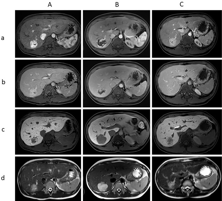
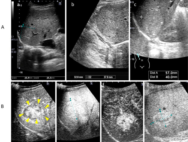
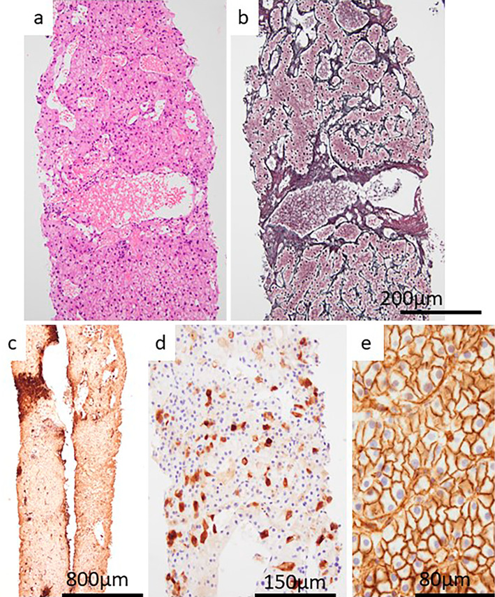
We reported a notable case of inflammatory hepatocellular adenoma that grew during pregnancy, consequently changing its appearance on magnetic resonance imaging remarkably. A 5-months-pregnant 35-year-old woman presented with a 37-mm liver nodule that had been diagnosed as focal nodular hyperplasia 3 years earlier. She had never used oral contraceptives. After 2 months, the nodule grew to 57 mm. The patient delivered a full-term infant without complications. Gadolinium-ethoxybenzyl-diethylenetriamine pentaacetic acid-enhanced magnetic resonance imaging performed after delivery revealed markedly different findings compared with the first images. A liver biopsy was performed, and the tumor was diagnosed as inflammatory hepatocellular adenoma.
Keywords: hepatocellular adenoma; magnetic resonance imaging; pregnancy; ultrasonography.
Conflict of interest statement
Figures
Similar articles
-
Growth of hepatocellular adenoma during pregnancy: A prospective study.J Hepatol. 2020 Jan;72(1):119-124. doi: 10.1016/j.jhep.2019.09.011. Epub 2019 Sep 21. J Hepatol. 2020. PMID: 31550458
-
Liver biopsy for diagnosis of presumed benign hepatocellular lesions lacking magnetic resonance imaging diagnostic features of focal nodular hyperplasia.Liver Int. 2016 Nov;36(11):1668-1676. doi: 10.1111/liv.13113. Epub 2016 Mar 30. Liver Int. 2016. PMID: 26969817
-
Hepatocellular adenoma(s) arising in nodular regenerative hyperplasia in a patient with systemic lupus erythematosus.Pathol Res Pract. 2020 Feb;216(2):152770. doi: 10.1016/j.prp.2019.152770. Epub 2019 Nov 28. Pathol Res Pract. 2020. PMID: 31810588
-
Focal Benign Liver Lesions and Their Diagnostic Pitfalls.Radiol Clin North Am. 2022 Sep;60(5):755-773. doi: 10.1016/j.rcl.2022.05.005. Epub 2022 Jul 4. Radiol Clin North Am. 2022. PMID: 35989043 Review.
-
Primary hepatocellular lesions: imaging findings on state-of-the-art magnetic resonance imaging, with pathologic correlation.Curr Probl Diagn Radiol. 2008 May-Jun;37(3):104-14. doi: 10.1067/j.cpradiol.2007.07.003. Curr Probl Diagn Radiol. 2008. PMID: 18436110 Review.
References
-
- Colombo M, Forner A, Ijzermans J, et al. . EASL clinical practice guidelines on the management of benign liver tumours. J Hepatol 65: 386-398, 2016. - PubMed
-
- Haring MPD, Spijkerboer CS, Cuperus FJC, et al. . Behavior and complications of hepatocellular adenoma during pregnancy and puerperium: a retrospective study and systematic review. HPB 23: 1152-1163, 2021. - PubMed
-
- Bioulac-Sage P, Gouw ASH, Balabaud C, Sempoux C. Hepatocellular adenoma: what we know, what we do not know, and why it matters. Histopathology 80: 878-897, 2022. - PubMed
-
- Dharmana H, Saravana-Bawan S, Girgis S, Low G. Hepatocellular adenoma: imaging review of the various molecular subtypes. Clin Radiol 72: 276-285, 2017. - PubMed
-
- Bonder A, Afdhal N. Evaluation of liver lesions. Clin Liver Dis 16: 271-283, 2012. - PubMed