

A cationic motif upstream Engrailed2 homeodomain controls cell internalization through selective interaction with heparan sulfates
- PMID: 37032404
- PMCID: PMC10083169
- DOI: 10.1038/s41467-023-37757-6
A cationic motif upstream Engrailed2 homeodomain controls cell internalization through selective interaction with heparan sulfates
Abstract
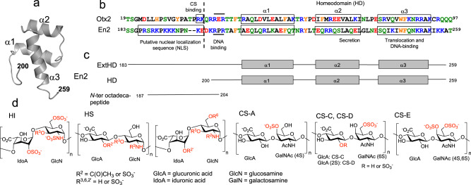
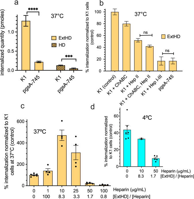
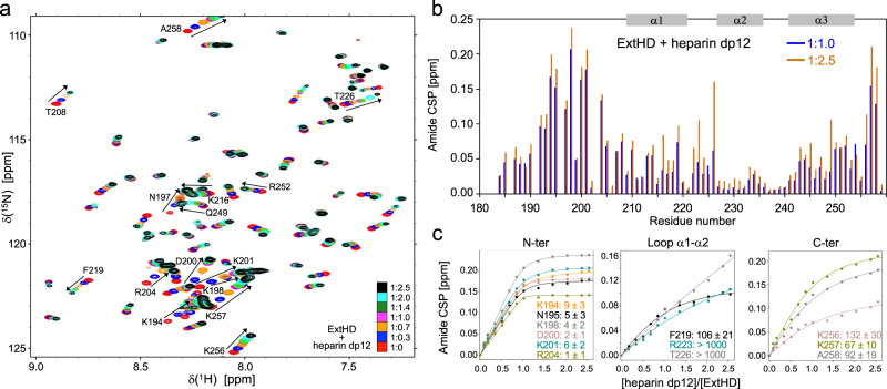
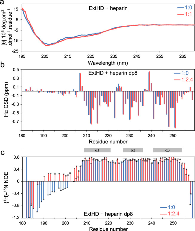
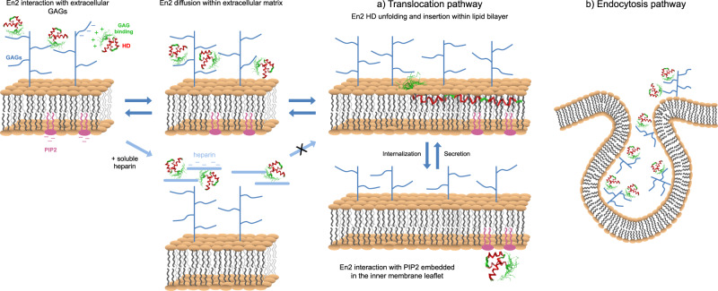
Engrailed2 (En2) is a transcription factor that transfers from cell to cell through unconventional pathways. The poorly understood internalization mechanism of this cationic protein is proposed to require an initial interaction with cell-surface glycosaminoglycans (GAGs). To decipher the role of GAGs in En2 internalization, we have quantified the entry of its homeodomain region in model cells that differ in their content in cell-surface GAGs. The binding specificity to GAGs and the influence of this interaction on the structure and dynamics of En2 was also investigated at the amino acid level. Our results show that a high-affinity GAG-binding sequence (RKPKKKNPNKEDKRPR), upstream of the homeodomain, controls En2 internalization through selective interactions with highly-sulfated heparan sulfate GAGs. Our data underline the functional importance of the intrinsically disordered basic region upstream of En2 internalization domain, and demonstrate the critical role of GAGs as an entry gate, finely tuning homeoprotein capacity to internalize into cells.
© 2023. The Author(s).
Conflict of interest statement
The authors declare no competing interests.
Figures
Similar articles
-
The B16F10 cell receptor for a metastasis-promoting site on laminin-1 is a heparan sulfate/chondroitin sulfate-containing proteoglycan.Cancer Res. 2002 Jun 15;62(12):3549-54. Cancer Res. 2002. PMID: 12068003
-
Cooperation of binding sites at the hydrophilic domain of cell-surface sulfatase Sulf1 allows for dynamic interaction of the enzyme with its substrate heparan sulfate.Biochim Biophys Acta. 2013 Nov;1830(11):5287-98. doi: 10.1016/j.bbagen.2013.07.014. Epub 2013 Jul 25. Biochim Biophys Acta. 2013. PMID: 23891937
-
Iduronic acid-containing glycosaminoglycans on target cells are required for efficient respiratory syncytial virus infection.Virology. 2000 Jun 5;271(2):264-75. doi: 10.1006/viro.2000.0293. Virology. 2000. PMID: 10860881
-
Sulfation of Glycosaminoglycans and Its Implications in Human Health and Disorders.Annu Rev Biomed Eng. 2017 Jun 21;19:1-26. doi: 10.1146/annurev-bioeng-071516-044610. Epub 2017 Feb 2. Annu Rev Biomed Eng. 2017. PMID: 28226217 Review.
-
The structure of glycosaminoglycans and their interactions with proteins.Chem Biol Drug Des. 2008 Dec;72(6):455-82. doi: 10.1111/j.1747-0285.2008.00741.x. Chem Biol Drug Des. 2008. PMID: 19090915 Review.
Cited by
-
ENGRAILED-1 transcription factor has a paracrine neurotrophic activity on adult spinal α-motoneurons.EMBO Rep. 2023 Aug 3;24(8):e56525. doi: 10.15252/embr.202256525. Epub 2023 Jul 6. EMBO Rep. 2023. PMID: 37534581 Free PMC article.
-
The limb dorsoventral axis: Lmx1b's role in development, pathology, evolution, and regeneration.Dev Dyn. 2024 Sep;253(9):798-814. doi: 10.1002/dvdy.695. Epub 2024 Jan 30. Dev Dyn. 2024. PMID: 38288855 Free PMC article. Review.
-
Translocation of penetratin-like peptides involving calcium-dependent interactions between glycosaminoglycans and phosphocholine headgroups of the membrane lipid bilayer.RSC Chem Biol. 2025 Aug 2;6(9):1391-402. doi: 10.1039/d5cb00099h. Online ahead of print. RSC Chem Biol. 2025. PMID: 40787615 Free PMC article.
References
-
- Dupont E, Prochiantz A, Joliot A. Identification of a signal peptide for unconventional secretion. J. Biol. Chem. 2007;282:8994–9000. - PubMed
-
- Spatazza J, et al. Homeoprotein signaling in development, health, and disease: a shaking of dogmas offers challenges and promises from bench to bed. Pharmacol. Rev. 2013;65:90–104. - PubMed
-
- Derossi D, Joliot AH, Chassaing G, Prochiantz A. The third helix of the Antennapedia homeodomain translocates through biological membranes. J. Biol. Chem. 1994;269:10444–10450. - PubMed
Publication types
MeSH terms
Substances
LinkOut - more resources
Full Text Sources
