. 2023 Mar 9;56(Pt 2):449-460.
doi: 10.1107/S1600576723001036. eCollection 2023 Apr 1.

A simple goniometer-compatible flow cell for serial synchrotron X-ray crystallography

Affiliations

A simple goniometer-compatible flow cell for serial synchrotron X-ray crystallography

Swagatha Ghosh et al. J Appl Crystallogr. .

Abstract

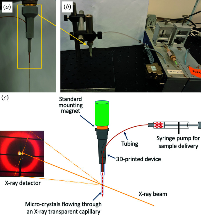
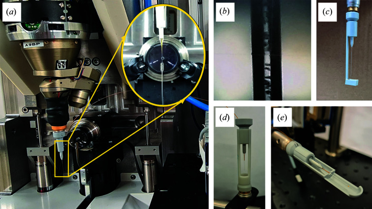
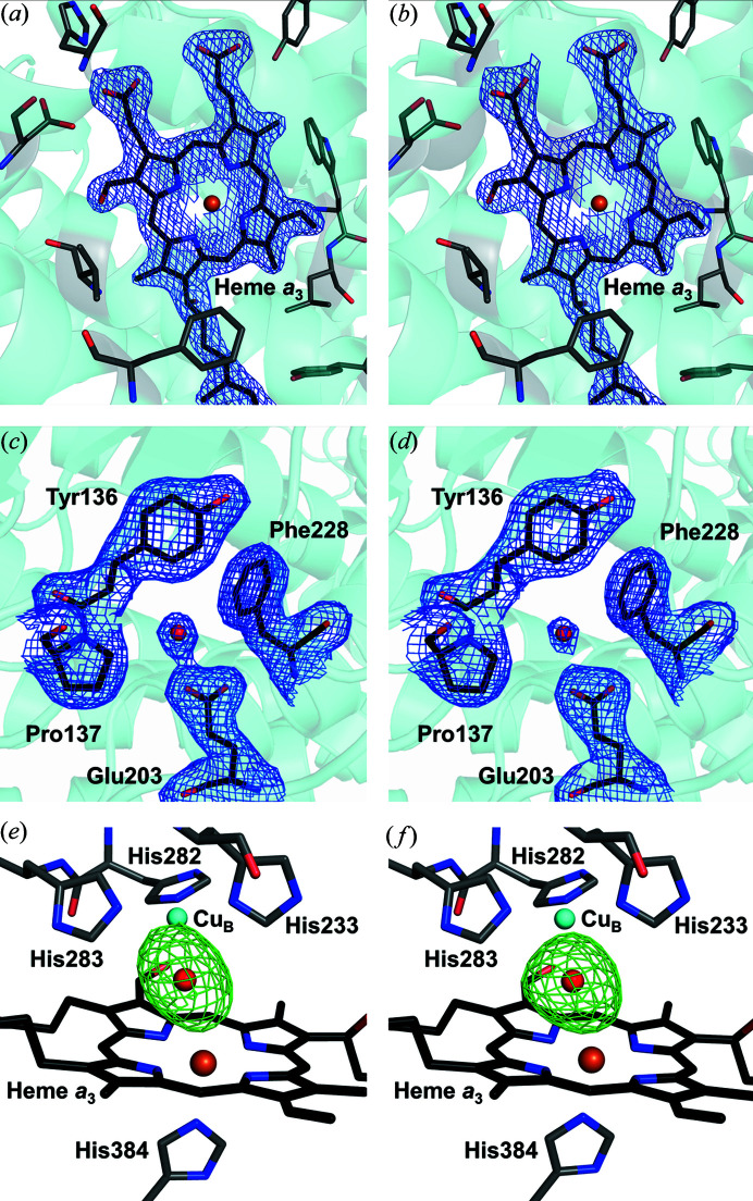
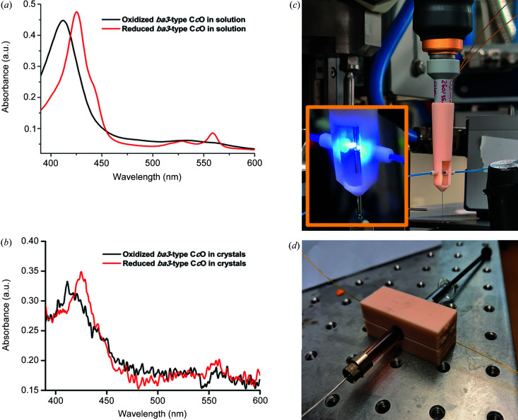
Serial femtosecond crystallography was initially developed for room-temperature X-ray diffraction studies of macromolecules at X-ray free electron lasers. When combined with tools that initiate biological reactions within microcrystals, time-resolved serial crystallography allows the study of structural changes that occur during an enzyme catalytic reaction. Serial synchrotron X-ray crystallography (SSX), which extends serial crystallography methods to synchrotron radiation sources, is expanding the scientific community using serial diffraction methods. This report presents a simple flow cell that can be used to deliver microcrystals across an X-ray beam during SSX studies. This device consists of an X-ray transparent glass capillary mounted on a goniometer-compatible 3D-printed support and is connected to a syringe pump via light-weight tubing. This flow cell is easily mounted and aligned, and it is disposable so can be rapidly replaced when blocked. This system was demonstrated by collecting SSX data at MAX IV Laboratory from microcrystals of the integral membrane protein cytochrome c oxidase from Thermus thermophilus, from which an X-ray structure was determined to 2.12 Å resolution. This simple SSX platform may help to lower entry barriers for non-expert users of SSX.

Keywords: cytochrome c oxidase; goniometer-compatible flow cells; macromolecular crystallography; serial synchrotron X-ray crystallography.


Figures

Figure 1
Figure 1
Overall design of the 3D-printed flow cell. (a) Flow cell with a 3D-printed base glued to an X-ray transparent glass (or quartz) capillary containing a channel through which a fused silica capillary tube is inserted and through which the sample is transported. This device is supported by a magnetic connection (either an iron or magnetized bullet) held by friction. The dimensions of the base can be adjusted to be compatible with any goniometer magnet used at any synchrotron radiation source. (b) Flow cell device connected to a syringe pump using suitable connectors. (c) Schematic of how the flow cell is used as a sample delivery system at a macromolecular crystallography beamline. Microcrystals (red) are injected from a syringe connected to a pump and delivered into an X-ray beam through an X-ray transparent capillary held in place on a standard goniometer magnet. X-ray diffraction data from microcrystals are collected on a rapid-readout X-ray detector.
Figure 2
Figure 2
Mounting and alignment of the 3D-printed flow cell on a macromolecular crystallography beamline. (a) Flow cell mounted on the goniometer magnet and aligned with the X-ray beam at BioMAX. (b) LCP crystals of ba 3-type CcO injected into the flow cell were observed through the glass capillary using the standard alignment optics of BioMAX. (c) Design of the catcher when the goniometer allows vertical mounting from above. This catcher is mounted by sliding over the flow cell and is held in place by friction. (d) Design of the catcher when the goniometer allows vertical mounting from below. In this case the sample also flows downwards. (e) Design of the catcher when the goniometer allows horizontal mounting.
Figure 3
Figure 3
Comparison between the electron density recovered from SSX studies using a flow cell and SFX studies using a high-viscosity injector. (a) 2F obs − F calc electron density map (blue, contoured at 1.5σ) showing SSX electron density for the active site heme a 3 of ba 3-type CcO determined at 2.12 Å resolution recovered using the flow cell for sample injection. (b) 2F obsF calc electron density map (blue, contoured at 1.5σ), showing SFX electron density for the active site heme a 3 at 2.3 Å resolution recovered using a high-viscosity injector for sample injection (Andersson et al., 2017 ▸). (c) 2F obsF calc electron density map (blue, contoured at 1.0σ) showing SSX electron density for a representative glutamic acid residue within the protein. (d) 2F obsF calc electron density map (blue, contoured at 1.0σ) showing SFX electron density for a representative glutamic acid residue within the protein. (e) F obsF calc omit electron density map (green, contoured at 4.5σ), showing a slightly elongated electron density in the active site of the SSX structure. (f) F obsF calc omit electron density map (green, contoured at 4.5σ) showing a slightly more spherical electron density in the active site of the SFX structure (Andersson et al., 2017 ▸). For both omit maps the electron density map is calculated without modelling any ligand between the heme a 3 iron and CuB.
Figure 4
Figure 4
Analysis of the contributions to background X-ray scattering when using 100 and 200 µm-diameter borosilicate glass capillaries. (a) Measured X-ray scattering, S(q), from empty 100 µm (blue) and 200 µm (red) diameter borosilicate glass capillaries after azimuthal integration. The X-ray scattering has arbitrary units (a.u.) and formula image , where 2θ is the angle of deflection of the X-rays and 1/d is the resolution quoted in X-ray crystallography, such that q = 2.0 Å−1 corresponds approximately to 3.1 Å resolution. (b) Measured X-ray scattering from a 100 µm-diameter flow cell containing LCP-grown microcrystals of CcO (mustard) and the best decomposition (black dashed line) of this scattering into its three scattering components from LCP (black solid line), borosilicate glass (blue) and air (black dotted line). (c) Measured X-ray scattering from a 200 µm-diameter flow cell containing LCP-grown microcrystals of CcO (mustard) and the best decomposition (black dashed line) of this scattering into its three scattering components of LCP (black solid line), borosilicate glass (red) and air (black dotted line). Air measurements were made without any object at the sample position, whereas LCP scattering was recorded from a homogeneous LCP sample extruded below the glass of a 200 µm-diameter flow cell. Air scattering was removed from the LCP and glass in panels (b) and (c). A larger background contribution from the borosilicate glass is observed when using the 100 µm-diameter capillary relative to the 200 µm glass capillary.
Figure 5
Figure 5
Spectroscopic measurements using the flow cell. (a) Absorbance spectra recorded from samples of purified ba 3-type CcO from T. thermophilus in solution. Red: CcO is purged of oxygen and reduced. Black: CcO in the oxidized state. (b) Absorbance spectra recorded from a slurry of CcO microcrystals held in a flow cell. Red: CcO is purged of oxygen and reduced. Black: CcO in the oxidized state. (c) Modified catcher, used to hold aligned optical fibres incoming from either side of the X-ray transparent capillary. This either allows absorption spectra to be recorded from samples as they pass by or allows light to be transported to the sample to activate photo-sensitive samples (inset). (d) Device for measuring absorption spectra from samples suspended in a glass syringe prior to injection using the flow cell. This is achieved using aligned optical fibres incoming from either side of the transparent syringe.

Similar articles

Cited by

References

    1. Andersson, R., Safari, C., Båth, P., Bosman, R., Shilova, A., Dahl, P., Ghosh, S., Dunge, A., Kjeldsen-Jensen, R., Nan, J., Shoeman, R. L., Kloos, M., Doak, R. B., Mueller, U., Neutze, R. & Brändén, G. (2019). Acta Cryst. D75, 937–946. - PMC - PubMed
    1. Andersson, R., Safari, C., Dods, R., Nango, E., Tanaka, R., Yamashita, A., Nakane, T., Tono, K., Joti, Y., Båth, P., Dunevall, E., Bosman, R., Nureki, O., Iwata, S., Neutze, R. & Brändén, G. (2017). Sci. Rep. 7, 4518. - PMC - PubMed
    1. Ballmoos, C. von, Gonska, N., Lachmann, P., Gennis, R. B., Ädelroth, P. & Brzezinski, P. (2015). Proc. Natl Acad. Sci. USA, 112, 3397–3402. - PMC - PubMed
    1. Basavappa, R., Petri, E. T. & Tolbert, B. S. (2003). J. Appl. Cryst. 36, 1297–1298.
    1. Beyerlein, K. R., Dierksmeyer, D., Mariani, V., Kuhn, M., Sarrou, I., Ottaviano, A., Awel, S., Knoska, J., Fuglerud, S., Jönsson, O., Stern, S., Wiedorn, M. O., Yefanov, O., Adriano, L., Bean, R., Burkhardt, A., Fischer, P., Heymann, M., Horke, D. A., Jungnickel, K. E. J., Kovaleva, E., Lorbeer, O., Metz, M., Meyer, J., Morgan, A., Pande, K., Panneerselvam, S., Seuring, C., Tolstikova, A., Lieske, J., Aplin, S., Roessle, M., White, T. A., Chapman, H. N., Meents, A. & Oberthuer, D. (2017). IUCrJ, 4, 769–777. - PMC - PubMed