. 2023 Mar 22:14:1138078.
doi: 10.3389/fendo.2023.1138078. eCollection 2023.

Vitamin D improves hepatic steatosis in NAFLD via regulation of fatty acid uptake and β-oxidation

Affiliations

Vitamin D improves hepatic steatosis in NAFLD via regulation of fatty acid uptake and β-oxidation

Tingwan Du et al. Front Endocrinol (Lausanne). .

Abstract

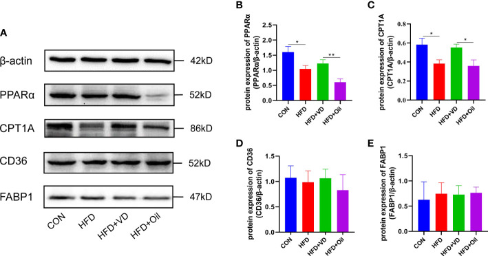
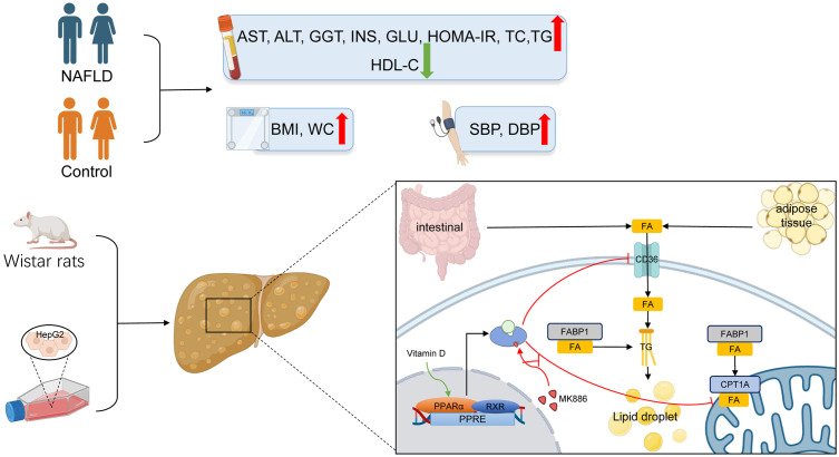
Introduction: The study aimed to explore the association of serum 25(OH)D3 and hepatic steatosis in non-alcoholic fatty liver disease (NAFLD) patients and to determine whether the effect of vitamin D (VD) is mediated by activation of the peroxisome proliferator-activated receptor α (PPARα) pathway.

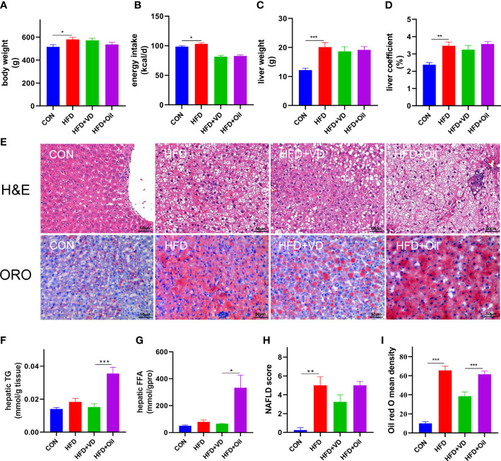
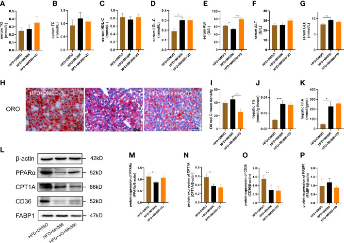
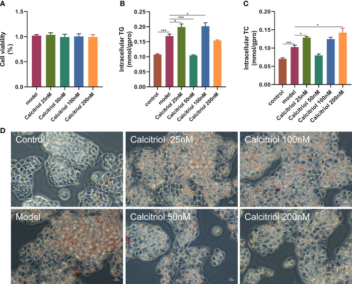
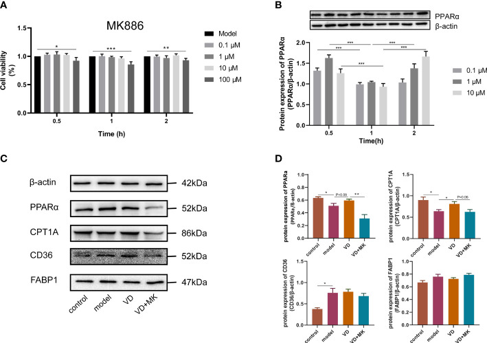
Methods: The study contained a case-control study, in vivo and in vitro experiments. A case-control study was conducted to compare serum parameters between NAFLD patients and controls and to evaluate the association of 25(OH)D3 and NAFLD. In vivo study, male Wistar rats were randomly divided into control and model groups, fed a standard chow diet and a high-fat diet (HFD), respectively, for 7 weeks to generate an NAFLD model. Then, the rats were treated with VD and a PPARα antagonist (MK886) for 7 weeks. Tissue and serum were collected and assessed by biochemical assays, morphological analysis, histological analysis, and western blot analysis. In vitro, HepG2 cells were incubated with oleic acid (OA) to induce steatosis, which was evaluated by staining. HepG2 cells were pretreated with MK886 followed by calcitriol treatment, and differences in lipid metabolism-related proteins were detected by western blot.

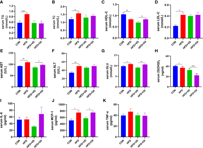
Results: NAFLD patients were characterized by impaired liver function, dyslipidemia, and insulin resistance. Serum 25(OH)D3 was negatively associated with alanine aminotransferase (ALT) in NAFLD. VD deficiency was a risk factor for patients with no advanced fibrosis. Adequate VD status (25(OH)D3 >20 ng/mL) had a protective effect in patients after adjustment for confounding variables. NAFLD rats showed hyperlipidemia with severe hepatic steatosis, systematic inflammation, and lower serum 25(OH)D3. VD treatment ameliorated hepatic steatosis both in NAFLD rats and OA-induced HepG2 cells. Further, MK886 inhibited the anti-steatosis effect of VD.

Conclusion: The study revealed that an adequate VD level may act as a protective factor in NAFLD and that VD may alleviate hepatic steatosis via the PPARα signaling pathway.

Keywords: PPARα; hepatic steatosis; lipid metabolism; non-alcoholic fatty liver disease; vitamin D.


Conflict of interest statement

The authors declare that the research was conducted in the absence of any commercial or financial relationships that could be construed as a potential conflict of interest.

Figures

Figure 1
Figure 1
Vitamin D treatment ameliorate hepatic steatosis in NAFLD rats. (A) body weight, (B) energy intake, (C)liver weight, (D) liver coefficient, (E) H&E staining of liver tissues (400 × magnification) and Oil red O staining of liver tissue (400 × magnification). Four rats in each group were captured three views for evaluation of steatosis, lobular inflammation and balloon degeneration. Both scores are examined by an experienced pathologist who was unaware of the study. (F) Quantification of hepatic TG and (G) FFA in liver tissue. (H) NAFLD score and (I) Oil red O density of liver. Data were shown as the mean ± SEM. (n=8); *P < 0.05, **P < 0.01, ***P < 0.001.
Figure 2
Figure 2
The effects of vitamin D in serum parameters of NAFLD rats. (A) serum TG, (B) TC, (C) HDL-C, (D) LDL-C, (E) AST, (F) ALT, (G) GLU, (H) 25(OH)D3, (I)IL-6, (J)MCP-1, (K)TNF-α levels. Data were shown as the mean ± SEM. (n=8) *P < 0.05, **P < 0.01, ***P < 0.001.
Figure 3
Figure 3
The effects of vitamin D on protein expression of lipid metabolism in NAFLD rats. (A) The expression of transcription factors involved in β-oxidation were detected by western blot. (B) PPARα and (C) CPT1A were qualified by β-actin. Lipid uptake proteins were detected by western blot. (D) CD36 and (E) FABP1 were qualified by β-actin. The band intensity ratios were analyzed by ImageJ. Data were shown as the mean ± SEM. (n=8) *P < 0.05, **P < 0.01.
Figure 4
Figure 4
MK886 inhibited the effect of Vitamin D3. (A) serum TG, (B) TC, (C) HDL-C, (D) LDL-C, (E) AST, (F) ALT, (G) GLU, (H) Oil red O staining of liver tissue (400 × magnification), (I) Oil red O mean density, (J) hepatic TG, and (K) FFA, (M–P) the fold change in expression of the proteins in (L) relative to the expression of b-actin. *P < 0.05, **P < 0.01, ***P < 0.001.
Figure 5
Figure 5
Effect of calcitriol on lipid deposition in HepG2 cells. Cytotoxic effects after (A) calcitriol (0-200nM) for 24 h were evaluated by CCK8 assay. (B) Intracellular TG and (C) TC were assayed in HepG2. (D) Intracellular lipid accumulation was detected by Oil red O staining method using an inverted microscope (400× magnification). Data were shown as the mean ± SEM. *P < 0.05, ***P < 0.001.
Figure 6
Figure 6
MK886 restrain the effect of VD treatment. (A) The cytotoxic effect after MK886 (0-100μM) for 0-2h was evaluated by CCK8 assay. (B) The best inhibiting effect after MK886 (0-10μM) for 0-2h was determined by western blot. (C, D) Effect of VD and MK886 on the protein expression involved in fatty acid uptake and β-oxidation, including PPARα, CPT1A, FABP1, CD36. The band intensity ratios were analyzed by ImageJ. Data are shown as means ± SEM. *P < 0.05, **P < 0.01, ***P < 0.001.
Figure 7
Figure 7
The lipid mechanism of NAFLD and anti-lipid accumulation mechanism of VD associated with PPARa pathway. Patients with NAFLD exhibited higher BMI, WC, SBP, DBP, AST, ALT, GGT, ALP, INS, GLU, HOMA-IR, TC, TG, LDL-C and lower HDL-C. The increased lipid oxidation and lipid uptake related proteins by VD treatment were inhibited after the application of MK886 in vivo and in vitro.

Similar articles

Cited by

References

    1. Machado MV, Cortez-Pinto H. NAFLD, MAFLD and obesity: brothers in arms? Nat Rev Gastroenterol Hepatol (2022) 20(2):67–8. doi: 10.1038/s41575-022-00717-4 - DOI - PubMed
    1. Lee CH, Lui DT, Lam KS. Non-alcoholic fatty liver disease and type 2 diabetes: An update. J Diabetes Invest (2022) 13(6):930–40. doi: 10.1111/jdi.13756 - DOI - PMC - PubMed
    1. Henderson GC. Plasma free fatty acid concentration as a modifiable risk factor for metabolic disease. Nutrients (2021) 13(8):2590. doi: 10.3390/nu13082590 - DOI - PMC - PubMed
    1. Khan RS, Bril F, Cusi K, Newsome PN. Modulation of insulin resistance in nonalcoholic fatty liver disease. Hepatol (Baltimore Md) (2019) 70(2):711–24. doi: 10.1002/hep.30429 - DOI - PubMed
    1. Milton-Laskibar I, Cuevas-Sierra A, Portillo MP, Martínez JA. Effects of resveratrol administration in liver injury prevention as induced by an obesogenic diet: Role of ruminococcaceae. Biomedicines (2022) 10(8):1797. doi: 10.3390/biomedicines10081797 - DOI - PMC - PubMed

Publication types