

Editorial
doi: 10.21037/tcr-22-2605.
Epub 2023 Feb 20.
Notes on the morphological features of cotyledonary dissecting leiomyoma, which is rare in clinical practice
Affiliations
- PMID: 37033355
- PMCID: PMC10080311
- DOI: 10.21037/tcr-22-2605
Item in Clipboard
Editorial
Notes on the morphological features of cotyledonary dissecting leiomyoma, which is rare in clinical practice
Transl Cancer Res.
.
No abstract available
Keywords: Cotyledonoid dissecting leiomyoma; leiomyoma; leiomyosarcoma; uterine mesenchymal tumor.
Conflict of interest statement
Conflicts of Interest: All authors have completed the ICMJE uniform disclosure form (available at https://tcr.amegroups.com/article/view/10.21037/tcr-22-2605/coif). The authors have no conflicts of interest to declare.
Figures
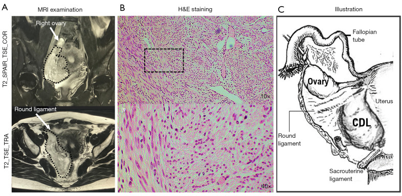
Morphological characteristics of a cotyledonoid dissecting leiomyoma observed on MRI and histopathological examination. (A) Results of MRI. The T2W1 image shows a continuous, low-to-high-intensity mass lesion originating within the right muscle layer of the uterine corpus, extending outward into the uterus. The mass (encircled by a dotted line) is present between the round ligament (white arrow) and uterine artery and is enclosed by a membrane. The right ovary (white arrow) present superior to the mass is normal. The images suggest that the mass may be a degenerative uterine leiomyoma growing from within the myometrium into the broad ligament. The leiomyoma measures approximately 20 mm in size on the posterior wall of the uterine fundus. No lesions are seen in the ovaries. Significant lymphadenopathy or ascites is absent. (B) Results of histopathological examination. A soft mass approximately 120 mm in size is seen protruding from the uterine serosal surface. The cut surface of the mass is grayish white and multinodular and demonstrates a proliferation of smooth muscle cells having an island-like/alveolar-like morphology with edematous stroma. Marked infiltration of these smooth muscle cells into the uterine smooth muscle layer is observed. There is also evidence of infiltration of some proliferating smooth muscle cells into the blood vessels. No severe nuclear atypia or mitotic cells are observed. On the basis of these findings, a diagnosis of cotyledonoid dissecting leiomyoma can be considered. Surgical pathological examination reveals no malignant findings in cervical and fallopian tube tissues. Upper panel: magnification ×10; lower panel: magnification ×40. (C) Illustration of the gross findings of the patient’s cotyledonoid dissecting leiomyoma. MRI, magnetic resonance imaging; H&E, hematoxylin and eosin; CDL, cotyledonoid dissecting leiomyoma.
Comment on
-
Case report: cotyledonoid dissecting leiomyoma in a 49-year-old woman.Transl Cancer Res. 2022 Nov;11(11):4189-4193. doi: 10.21037/tcr-22-1521. Transl Cancer Res. 2022. PMID: 36523320 Free PMC article.
Similar articles
-
An unusual case of uterine cotyledonoid dissecting leiomyoma with adenomyosis.Diagn Pathol. 2016 Aug 4;11(1):69. doi: 10.1186/s13000-016-0523-1. Diagn Pathol. 2016. PMID: 27491369 Free PMC article.
-
Cotyledonoid Dissecting Leiomyoma: A Rare Variant of Leiomyoma of the Uterus.Cureus. 2022 Oct 16;14(10):e30352. doi: 10.7759/cureus.30352. eCollection 2022 Oct. Cureus. 2022. PMID: 36407217 Free PMC article.
-
Cotyledonoid dissecting leiomyoma: an uncommon form of a common disease.Obstet Gynecol Sci. 2019 Sep;62(5):362-366. doi: 10.5468/ogs.2019.62.5.362. Epub 2019 Jul 26. Obstet Gynecol Sci. 2019. PMID: 31538081 Free PMC article.
-
Cotyledonoid Leiomyoma Clinical Characteristics, Imaging Features, and Review of the Literature.J Ultrasound Med. 2021 Jul;40(7):1459-1469. doi: 10.1002/jum.15510. Epub 2020 Sep 21. J Ultrasound Med. 2021. PMID: 32955750 Review.
-
Cotyledonoid leiomyoma: a benign uterine tumor with alarming gross appearance.Arch Pathol Lab Med. 2002 Feb;126(2):210-3. doi: 10.5858/2002-126-0210-CL. Arch Pathol Lab Med. 2002. PMID: 11825122 Review.
References
-
- Uterine leiomyoma. Female Genital Tumours WHO Classification of Tumours, 5th ed., Vol.4. WHO Classification of Tumours Editorial Board. World Health Organization. 2020:272-6.
-
- WHO classification of mesenchymal tumours of the lower genital tract. Female Genital Tumours WHO Classification of Tumours, 5th ed., Vol.4. WHO Classification of Tumours Editorial Board. World Health Organization. 2020:13.
-
- Zaloudek CJ, Hendrickson MR, Soslow RA. Mesenchymal tumors of the uterus. In Kurman R, editor. Blaustein’s Pathology of the Female Genital Tract, 6th ed. Springer, New York, 2010:453-527.
-
- Intravenous leiomyotosis. Female Genital Tumours WHO Classification of Tumours, 5th ed., Vol.4. WHO Classification of Tumours Editorial Board. World Health Organization. 2020:277-8.
Publication types
LinkOut - more resources
Full Text Sources