. 2023 Mar 9;14(14):1591-1601.
doi: 10.1039/d3py00075c. eCollection 2023 Apr 4.

Nucleophile responsive charge-reversing polycations for pDNA transfection

Affiliations

Nucleophile responsive charge-reversing polycations for pDNA transfection

Reece W Lewis et al. Polym Chem. .

Abstract

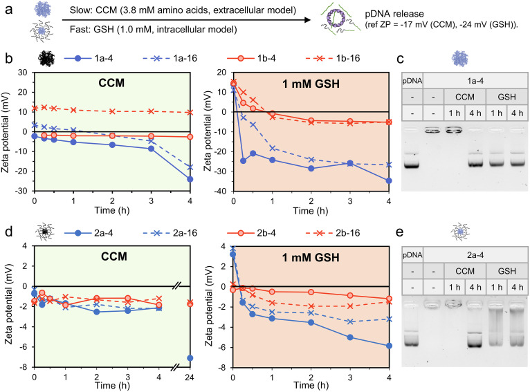
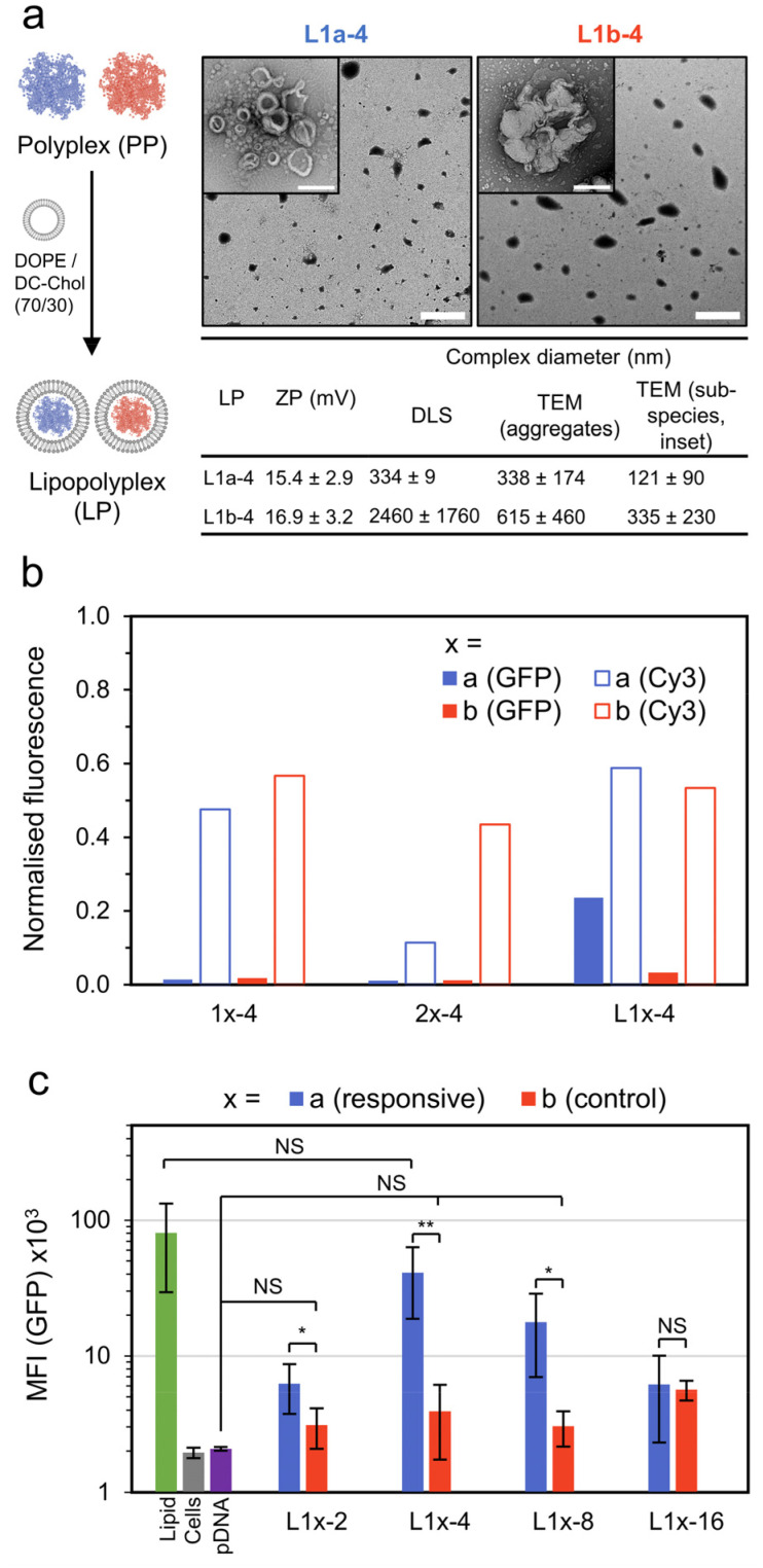
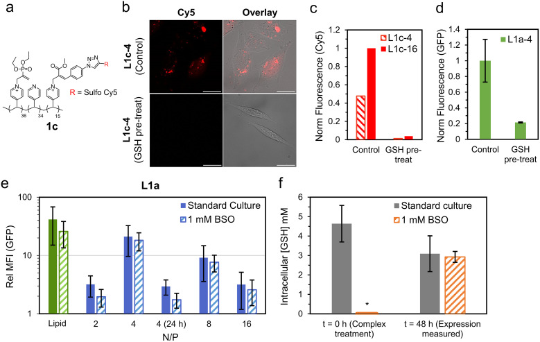
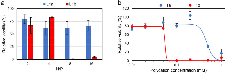
Polycationic carriers promise low cost and scalable gene therapy treatments, however inefficient intracellular unpacking of the genetic cargo has limited transfection efficiency. Charge-reversing polycations, which transition from cationic to neutral or negative charge, can offer targeted intracellular DNA release. We describe a new class of charge-reversing polycation which undergoes a cationic-to-neutral conversion by a reaction with cellular nucleophiles. The deionization reaction is relatively slow with primary amines, and much faster with thiols. In mammalian cells, the intracellular environment has elevated concentrations of amino acids (∼10×) and the thiol glutathione (∼1000×). We propose this allows for decationization of the polymeric carrier slowly in the extracellular space and then rapidly in the intracellular milleu for DNA release. We demonstrate that in a lipopolyplex formulation this leads to both improved transfection and reduced cytotoxicity when compared to a non-responsive polycationic control.


Conflict of interest statement

There are no conflicts of interest.

Figures

Fig. 1
Fig. 1. Outline of nucleophile responsive charge-reversing polycation study. (a) General structure of responsive polycation and its charge-reversing reaction with S/N-nucleophiles. Representative 1H NMR conversion data for the decationization reaction in phosphate buffer (pH 7.4, 100 mM) at RT ([cationic units] = [nucleophile] = 7 mM), see Fig. S1 and S2 for additional data. Curves are fit with pseudo first-order kinetic model. (b) General structure of non-responsive polycation. (c) Scheme showing proposed two-stage charge reversing process of responsive pDNA complexes in extracellular and intracellular conditions. *In this work cellular transfection is demonstrated by combining the polymer complexes with a lipid.
Fig. 2
Fig. 2. Preparation of polycations and their pDNA (pEGFP-C1) complexes. (a) Preparation of polyamines by RAFT polymerisation followed by their reaction with electrophiles to generate either responsive or permanently cationic (control) polycations. (b) Formation of micelle and polyplex type pDNA complexes studied in this work. (c) Titration of polycations to pDNA (6.5 μg mL−1 pDNA; 20 μM phosphates) as a function of N/P (prepared in 10 mM pH 7.4 phosphate buffer). (d) Summary of N/P = 4 pDNA complex size. Diameters are reported as the average across 3 samples (DLS) or 3 images (TEM) ± SD. For TEM, diameter is the length of the longest dimension. (e) TEM images of N/P = 4 pDNA complexes (stained, 2 wt% uranyl acetate), scale bar 200 nm. Additional characterisation data can be found in Fig. S4–14 (gel electrophoresis, polymer characterisation and additional DLS/TEM data).
Fig. 3
Fig. 3. Decationization and pDNA release studies under model intra- and extra-cellular conditions. Samples were maintained at 37 °C and the concentration of pDNA (as anionic phosphates) was maintained at 0.02 mM for zeta potential (ZP) measurements and 0.06 mM for gel electrophoresis. (a) Expected responsive complex pDNA release under model extracellular (HAMS-F12 media, CCM) and intracellular (1.0 mM GSH) conditions. (b) Change in ZP over time for responsive (blue) and non-responsive (red) polyplexes in CCM and 1 mM GSH. (c) Gel electrophoresis measurements of 1a-4 after incubation in CCM or 1 mM GSH for 1 and 4 h. (d) Change in ZP over time for responsive (blue) and non-responsive (red) micelles in CCM and 1 mM GSH. (e) Gel electrophoresis measurements of 2a-4 after incubation in CCM or 1 mM GSH for 1 and 4 h. Additional gel electrophoresis data in Fig. S15 and S16.
Fig. 4
Fig. 4. Lipopolyplex preparation, EGFP transfection and pDNA localisation data. (a) Preparation and particle characterisation of responsive and control lipopolyplexes (lipid : pDNA = 10 : 1 w/w). Lipopolyplexes are composed of aggregates (main TEM image, scale bar 2 μm) of smaller sub-species (inset image, scale bar 200 nm). (b) Normalized fluorescence intensity of CHO cells treated with various pDNA (1 μg per well) complexes as measured by CLSM. GFP fluorescence measured 48 h after treatment and Cy3 fluorescence (from Cy3 labelled pDNA, λex = 543 nm, λdet = 548–797 nm) measured 4 h after treatment. The results are normalised to a lipid formulation (DOPE/DC-Chol : 70/30). (c) Mean GFP fluorescence intensity (MFI) of CHO cells 48 h after treatment with control and responsive lipopolyplexes as measured by flow cytometry (n = 5, each measurement >10k cells, error bars are ±SD, NS = p > 0.05, * = p < 0.05, ** = p < 0.01). Additional TEM and CLSM images in Fig. S17–S21.
Fig. 5
Fig. 5. Investigations into role of nucleophile responsive polycationic functionality. (a) Polymer structure of 1c, a Cy5 labelled responsive polycation. (b) Cy5 (red) localisation study of CHO cells treated with lipopolyplexes prepared using 1c (L1c-4, Control). These are compared to cells treated with matching lipopolyplexes prepared from 1c pre-treated with 5× excess GSH (L1c-4, GSH pre-treat). Images were taken using CLSM with Cy5 (λex = 633 nm, λdet = 638–797 nm, red) and bright field channels overlayed, scale bar is 25 μm. (c) Normalised Cy5 fluorescence of cells treated with lipopolyplexes prepared from 1c (Control) and decationized 1c (GSH pre-treat). (d) Normalised EGFP transfection of cells treated with L1a-4 (Control) compared to L1a-4 with GSH pre-treatment (2 mM GSH in CCM, 37 °C for 1 h). Data is mean ± SD (n = 3) as evaluated by flow cytometry. (e) GFP fluorescence 48 h (or 24 h where stated) of lipopolyplex treated CHO cells relative to untreated cells under the same culture condition (standard culture or 1 mM BSO). Data is mean ± SD (n ≥ 3) as evaluated by flow cytometry. (f) Intracellular GSH concentration of standard culture and BSO treated cells at t = 0 and 48 h. Data is mean ± SD (n = 4) as measured using a commercial luminescence-based assay which produces luciferase in the presence of GSH (Fig. S28†). *For t = 0 (1 mM BSO) intracellular GSH was below limit of detection.
Fig. 6
Fig. 6. In vitro cytotoxicity studies on CHO cells via MTS assay (mean ± SD, n = 3). Cells were treated with (a) lipopolyplexes (0.5 μg pDNA per well) at various N/P ratios and (b) responsive and non-responsive polycations. Data is fitted using sigmoidal four-parameter logistic model, with EC50 determined as 0.47 mM for 1a and 0.05 mM for 1b, see Fig. S32 for full description of fitting parameters. Note: at N/P = 4 lipopolyplexes contain a polycation concentration of 0.06 mM.

Similar articles

Cited by

References

    1. Alnasser S. M. Gene. 2021;769:145246. doi: 10.1016/j.gene.2020.145246. - DOI - PubMed
    1. Uchida S. Kataoka K. J. Biomed. Mater. Res., Part A. 2019;107:978–990. doi: 10.1002/jbm.a.36614. - DOI - PubMed
    1. Muhammad K. Zhao J. Gao B. Feng Y. J. Mater. Chem. B. 2020;8:9621–9641. doi: 10.1039/D0TB01675F. - DOI - PubMed
    1. Kumar R. Santa Chalarca C. F. Bockman M. R. Bruggen C. V. Grimme C. J. Dalal R. J. Hanson M. G. Hexum J. K. Reineke T. M. Chem. Rev. 2021;121:11527–11652. doi: 10.1021/acs.chemrev.0c00997. - DOI - PubMed
    1. Dutta K. Das R. Medeiros J. Kanjilal P. Thayumanavan S. Adv. Funct. Mater. 2021;31:2011103. doi: 10.1002/adfm.202011103. - DOI - PMC - PubMed

LinkOut - more resources