. 2023 Mar 22:14:1130735.
doi: 10.3389/fimmu.2023.1130735. eCollection 2023.

Pharmacological depletion of microglia alleviates neuronal and vascular damage in the diabetic CX3CR1-WT retina but not in CX3CR1-KO or hCX3CR1I249/M280-expressing retina

Affiliations

Pharmacological depletion of microglia alleviates neuronal and vascular damage in the diabetic CX3CR1-WT retina but not in CX3CR1-KO or hCX3CR1I249/M280-expressing retina

Kaira A Church et al. Front Immunol. .

Abstract

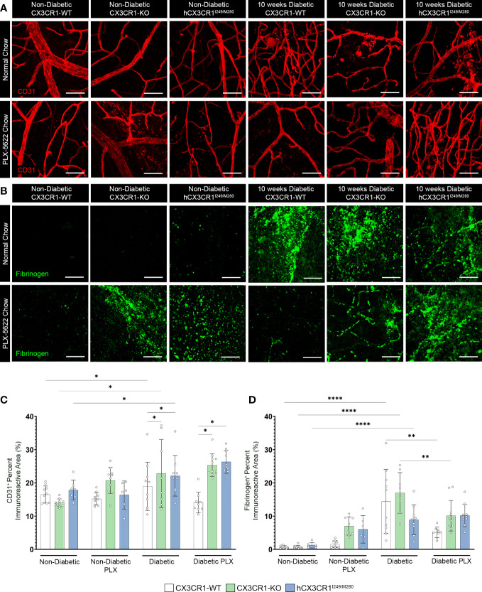
Diabetic retinopathy, a microvascular disease characterized by irreparable vascular damage, neurodegeneration and neuroinflammation, is a leading complication of diabetes mellitus. There is no cure for DR, and medical interventions marginally slow the progression of disease. Microglia-mediated inflammation in the diabetic retina is regulated via CX3CR1-FKN signaling, where FKN serves as a calming signal for microglial activation in several neuroinflammatory models. Polymorphic variants of CX3CR1, hCX3CR1I249/M280 , found in 25% of the human population, result in a receptor with lower binding affinity for FKN. Furthermore, disrupted CX3CR1-FKN signaling in CX3CR1-KO and FKN-KO mice leads to exacerbated microglial activation, robust neuronal cell loss and substantial vascular damage in the diabetic retina. Thus, studies to characterize the effects of hCX3CR1I249/M280 -expression in microglia-mediated inflammation in the diseased retina are relevant to identify mechanisms by which microglia contribute to disease progression. Our results show that hCX3CR1I249/M280 mice are significantly more susceptible to microgliosis and production of Cxcl10 and TNFα under acute inflammatory conditions. Inflammation is exacerbated under diabetic conditions and coincides with robust neuronal loss in comparison to CX3CR1-WT mice. Therefore, to further investigate the role of hCX3CR1I249/M280 -expression in microglial responses, we pharmacologically depleted microglia using PLX-5622, a CSF-1R antagonist. PLX-5622 treatment led to a robust (~70%) reduction in Iba1+ microglia in all non-diabetic and diabetic mice. CSF-1R antagonism in diabetic CX3CR1-WT prevented TUJ1+ axonal loss, angiogenesis and fibrinogen deposition. In contrast, PLX-5622 microglia depletion in CX3CR1-KO and hCX3CR1I249/M280 mice did not alleviate TUJ1+ axonal loss or angiogenesis. Interestingly, PLX-5622 treatment reduced fibrinogen deposition in CX3CR1-KO mice but not in hCX3CR1I249/M280 mice, suggesting that hCX3CR1I249/M280 expressing microglia influences vascular pathology differently compared to CX3CR1-KO microglia. Currently CX3CR1-KO mice are the most commonly used strain to investigate CX3CR1-FKN signaling effects on microglia-mediated inflammation and the results in this study indicate that hCX3CR1I249/M280 receptor variants may serve as a complementary model to study dysregulated CX3CR1-FKN signaling. In summary, the protective effects of microglia depletion is CX3CR1-dependent as microglia depletion in CX3CR1-KO and hCX3CR1I249/M280 mice did not alleviate retinal degeneration nor microglial morphological activation as observed in CX3CR1-WT mice.

Keywords: CX3CR1 chemokine receptor; depletion; diabetic retinopathy; inflammation; microglia.


Conflict of interest statement

Author SG was employed by Biogen, Cambridge, MA, and is currently employed by Cerevel Therapeutics, Cambridge, MA. Author ACC is employed full-time by Biogen, Cambridge, MA. Author RR was employed full-time by Biogen, Cambridge, MA, and currently employed full-time by Third Rock Ventures, Boston, MA. The remaining authors declare that the research was conducted in the absence of any commercial or financial relationships that could be construed as a potential conflict of interest.

Figures

Figure 1
Figure 1
Acute LPS-induced inflammation induces aberrant angiogenesis and gliosis in the hCX3CR1I249/M280 retina. hCX3CR1I249/M280 mice were i.p. injected with 0.08mg/Kg LPS once daily for four days (4d), and naïve age-matched controls received PBS. (A) Confocal images of retinal tissues stained for Iba1 (green), CD31 (Red), GFAP (magenta) and DAPI (blue) in naïve and 4d LPS hCX3CR1I249/M280 mice. Confocal images represent the retinal ganglion cell layer in the peripheral retina. (B–D), Quantification of retinal IHC analysis for Iba1+ cells/mm3 (B), CD31+ percent immunoreactive area (C), and percent immunoreactive area for GFAP (D). (E–G), Graphical representation for RT-qPCR analysis of retinas for relative mRNA expression for Cxcl10 (E), Tnfα (F) and Il10 (G). Data show the average of the 2 central, 2 medial and 2 peripheral images taken per mouse. Data show mean ± SD, n = 3 to 10 mice per group where each dot represents an individual mouse. **P<0.01, ***P<0.001, ****P<0.0001 using Student’s t-test, with Welch’s correction. Scale bars measure 50µm.
Figure 2
Figure 2
hCX3CR1I249/M280 variant is associated with increased neuronal cell loss and reactive gliosis in the diabetic retina. Hyperglycemia was induced in CX3CR1-WT and hCX3CR1I249/M280 mice via i.p. injection with STZ and retinal tissues were analyzed after four months of hyperglycemia. (A) Confocal images of retinal tissues stained for Iba1 (green), NeuN (Red) and GFAP (magenta) in naïve CX3CR1-WT and 4-months diabetic CX3CR1-WT and hCX3CR1I249/M280 mice. Confocal images represent the retinal ganglion cell layer in the peripheral retina. B-D, Quantification of retinal IHC analysis for NeuN+ cells/mm3 (B), Iba1+ cells/mm3 (C), and percent immunoreactive area for GFAP (D). Data show the 2 central, 2 medial and 2 peripheral images taken per mouse. Data show mean ± SD, n = 4 to 10 mice per group where each dot represents an individual mouse. *P<0.05, **P<0.01, ***P<0.001 ****P<0.0001 using Student’s t-test, with Welch’s correction. Scale bars measure 50µm.
Figure 3
Figure 3
CSF-1R antagonism induces robust microglia depletion in the diabetic retina. (A) Microglia were pharmacologically depleted using PLX-5622 in non-diabetic and 8-wks diabetic CX3CR1-WT, CX3CR1-KO, and hCX3CR1I249/M280 mice for two weeks. Non-diabetic control mice received citrate buffer. Non-depleted, non-diabetic and diabetic controls remained on normal chow. Confocal images of retinal tissues stained for Iba1 (green) (B) and transformation index cellular tracings (C) in CX3CR1-WT, CX3CR1-KO and hCX3CR1I249/M280 mice. Confocal images represent the retinal ganglion cell layer in the peripheral retina. (D, E), Quantification of retinal IHC analysis for Iba1+ cells/mm3 (D) and transformation index (E). Data show the average of the 2 central, 2 medial and 2 peripheral images taken per mouse. Data show mean ± SD, n = 6 to 10 mice per group where each dot represents an individual mouse (D). Transformation index data show mean ± SD n = 44 to 75 microglia per group for n=5 mice where each dot represents an individual microglia cell (E). ****P<0.0001 using 2-way ANOVA. Scale bars measure 50µm.
Figure 4
Figure 4
PLX-5622 treatment does not prevent TUJ1+ axonal loss in the diabetic CX3CR1-KO and hCX3CR1I249/M280 retinas. Microglia were pharmacologically depleted using PLX-5622 in non-diabetic and 8-wks diabetic CX3CR1-WT, CX3CR1-KO, and hCX3CR1I249/M280 mice for two weeks. Non-diabetic control mice received citrate buffer. Non-depleted, non-diabetic and diabetic controls remained on normal chow. Confocal images of retinal tissues stained for TUJ1 (turquoise) (A) and GFAP (magenta) (B) in CX3CR1-WT, CX3CR1-KO and hCX3CR1I249/M280 mice. Confocal images represent the retinal ganglion cell layer in the peripheral retina. (C, D), Quantification of retinal IHC analysis for TUJ1+ percent immunoreactive area (C) and GFAP+ percent immunoreactive area (D). Data show the average of the 2 central, 2 medial and 2 peripheral images taken per mouse. Data show mean ± SD, n = 6 to 10 mice per group where each dot represents an individual mouse. **P<0.01, ***P<0.001, ****P<0.0001 using 2-way ANOVA. Scale bars measure 50µm.
Figure 5
Figure 5
PLX-5622 treatment does not alleviate angiogenesis in the diabetic CX3CR1-KO and hCX3CR1I249/M280 retina. Microglia were pharmacologically depleted using PLX-5622 in non-diabetic and 8-wks diabetic CX3CR1-WT, CX3CR1-KO, and hCX3CR1I249/M280 mice for two weeks. Non-diabetic control mice received citrate buffer. Non-depleted, non-diabetic and diabetic controls remained on normal chow. Confocal images of retinal tissues stained for CD31 (red) (A) and fibrinogen (green) (B) in CX3CR1-WT, CX3CR1-KO and hCX3CR1I249/M280 mice. Confocal images represent the retinal ganglion cell layer in the peripheral retina. (C, D), Quantification of retinal IHC analysis for CD31+ percent immunoreactive area (C) and fibrinogen+ percent immunoreactive area (D). Data show the average of the 2 central, 2 medial and 2 peripheral images taken per mouse. Data show mean ± SD, n = 6 to 10 mice per group where each dot represents an individual mouse. *P<0.05, **P<0.01, ****P<0.0001 using 2-way ANOVA. Scale bars measure 50µm.
Figure 6
Figure 6
Schematic summary: Pharmacological depletion of microglia alleviates neuronal and vascular damage in the diabetic CX3CR1-WT retina but not in CX3CR1-KO or hCX3CR1I249/M280 -expressing retina. In the non-diabetic retina, normal chow and PLX-5622 chow treated CX3CR1-WT, CX3CR1-KO, and hCX3CR1I249/M280 mice have intact vasculature and ramified microglia. PLX-5622 chow treatment induces robust microglia depletion in CX3CR1-WT, CX3CR1-KO, and hCX3CR1I249/M280 mice. Prolonged hyperglycemia results in angiogenesis, vascular damage and fibrinogen deposition in CX3CR1-WT, CX3CR1-KO, and hCX3CR1I249/M280 mice. Additionally, diabetic CX3CR1-KO, and hCX3CR1I249/M280 mice are more susceptible to neuronal and axonal loss. CSF-1R antagonism alleviates vascular damage and fibrinogen deposition and prevents neuronal loss in the diabetic CX3CR1-WT retina. In contrast, CSF-1R antagonism in CX3CR1-KO, and hCX3CR1I249/M280 mice does not prevent vascular damage, fibrinogen deposition and neuronal loss.

References

    1. Cecilia OM, José Alberto CG, José NP, Ernesto Germán CM, Ana Karen LC, Luis Miguel RP, et al. . Oxidative stress as the main target in diabetic retinopathy pathophysiology. J Diabetes Res (2019) 2019:8562408. doi: 10.1155/2019/8562408 - DOI - PMC - PubMed
    1. Ren J, Zhang S, Pan Y, Jin M, Li J, Luo Y, et al. . Diabetic retinopathy: Involved cells, biomarkers, and treatments. Front Pharmacol (2022) 13:953691. doi: 10.3389/fphar.2022.953691 - DOI - PMC - PubMed
    1. McLeod D. Why cotton wool spots should not be regarded as retinal nerve fibre layer infarcts. Br J Ophthalmol (2005) 89(2):229–37. doi: 10.1136/bjo.2004.058347 - DOI - PMC - PubMed
    1. Wang W, Lo ACY. Diabetic retinopathy: Pathophysiology and treatments. Int J Mol Sci (2018) 19(6). doi: 10.3390/ijms19061816 - DOI - PMC - PubMed
    1. Ibrahim AS, El-Shishtawy MM, Peña A, Jr., Liou GI. Genistein attenuates retinal inflammation associated with diabetes by targeting of microglial activation. Mol Vis (2010) 16:2033–42. Available at: https://www.molvis.org/molvis/v16/a219/. - PMC - PubMed

Publication types