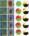
Detecting changes in the blood flow of the optic disk in patients with nonarteritic anterior ischemic optic neuropathy via optical coherence tomography-angiography
- PMID: 37034068
- PMCID: PMC10081673
- DOI: 10.3389/fneur.2023.1140770
Detecting changes in the blood flow of the optic disk in patients with nonarteritic anterior ischemic optic neuropathy via optical coherence tomography-angiography
Abstract
Purpose: This study aimed to evaluate the changes in the blood flow of the optic disk in patients with nonarteritic anterior ischemic optic neuropathy (NAION) using optical coherence tomography-angiography (OCTA) and to investigate the relationship among the changes in the blood flow of the optic disk, visual field defect, peripapillary retinal nerve fiber layer (RNFL), and macular ganglion cell complex (mGCC).
Methods: This was a prospective observational case series study. A total of 89 patients (89 eyes) with NAION were included in this study. All patients underwent best corrected visual acuity (BCVA), slit-lamp and direct ophthalmoscopic examinations, color fundus photography, visual field test, and blood flow indicators of the radial peripapillary capillaries (RPC) including whole en face image vessel density (VD), peripapillary VD by OCTA, the peripapillary RNFL, and mGCC by spectral-domain optic coherence tomography (OCT). The changes of blood flow in the optic disk at ≤3, 4-8, 9-12, 13-24, and >24 weeks of the natural course of NAION were measured, and the relationship among the changes in the blood flow of the optic disk, visual field defect, peripapillary RNFL, and mGCC was also analyzed.
Results: The mean age of 89 patients with NAION was 56.42 ± 6.81 years (ranging from 39 to 79). The initial RPC whole en face image VD was significantly reduced after acute NAION (≤3 weeks) (F = 45.598, P < 0.001) and stabilized from the eighth week onward. Over the course of NAION, the superonasal RPC, superior mGCC, and superotemporal RNFL decreased mostly with time (F = 95.658, 109.787, 263.872, respectively; P < 0.001). Maximal correlations were found between superior mGCC and temporosuperior RPC in the NAION phase (R = 0.683, P < 0.01) and between superonasal RPC and superonasal RNFL (R = 0.740, P < 0.01). The mean defect was correlated with temporosuperior RPC (R = -0.281, P < 0.01) and superior mGCC (R = -0.160, P = 0.012).
Conclusion: Over the course of NAION, OCTA shows a tendency toward change in the retinal capillary plexus of the optic disk. OCTA is proved to be a practical and useful tool for observing papillary perfusion in NAION.
Keywords: ganglion cell complex (GCC); nonarteritic anterior ischaemic optic neuropathy; optical coherence tomography angiography; vessel density (VD); visual field (VF).
Copyright © 2023 Xiao and Sun.
Conflict of interest statement
The authors declare that the research was conducted in the absence of any commercial or financial relationships that could be construed as a potential conflict of interest.
Figures

Similar articles
-
Optical coherence tomography angiography of peripapillary vessel density in non-arteritic anterior ischemic optic neuropathy and demyelinating optic neuritis.Front Neurol. 2024 Oct 29;15:1432753. doi: 10.3389/fneur.2024.1432753. eCollection 2024. Front Neurol. 2024. PMID: 39539649 Free PMC article.
-
Follow-Up of Nonarteritic Anterior Ischemic Optic Neuropathy With Optical Coherence Tomography Angiography.Invest Ophthalmol Vis Sci. 2021 Feb 1;62(2):42. doi: 10.1167/iovs.62.4.42. Invest Ophthalmol Vis Sci. 2021. PMID: 33635311 Free PMC article.
-
Retinal and Choroidal Microvasculature in Nonarteritic Anterior Ischemic Optic Neuropathy: An Optical Coherence Tomography Angiography Study.Invest Ophthalmol Vis Sci. 2018 Feb 1;59(2):870-877. doi: 10.1167/iovs.17-22996. Invest Ophthalmol Vis Sci. 2018. PMID: 29490340
-
Microvascular alterations detected by optical coherence tomography angiography in non-arteritic anterior ischaemic optic neuropathy: a meta-analysis.Acta Ophthalmol. 2022 Mar;100(2):e386-e395. doi: 10.1111/aos.14930. Epub 2021 Jun 21. Acta Ophthalmol. 2022. PMID: 34155823
-
[Nonarteritic ischemic optic neuropathy animal model and its treatment applications].Nippon Ganka Gakkai Zasshi. 2014 Apr;118(4):331-61. Nippon Ganka Gakkai Zasshi. 2014. PMID: 24864434 Review. Japanese.
Cited by
-
Optical coherence tomography angiography of peripapillary vessel density in non-arteritic anterior ischemic optic neuropathy and demyelinating optic neuritis.Front Neurol. 2024 Oct 29;15:1432753. doi: 10.3389/fneur.2024.1432753. eCollection 2024. Front Neurol. 2024. PMID: 39539649 Free PMC article.
References
-
- Raizada K, Margolin E. Non-arteritic Anterior Ischemic Optic Neuropathy. Treasure Island, FL: StatPearls Publishing; (2022). - PubMed
-
- van Oterendorp C, Lagrèze WA, Feltgen N. Pathogenese und therapie der nicht arteriitischen anterioren ischämischen optikusneuropathie [non-arteritic anterior ischaemic optic neuropathy: pathogenesis and therapeutic approaches]. Klin Monbl Augenheilkd. (2019) 236:1283–91. 10.1055/a-0972-1625 - DOI - PubMed
LinkOut - more resources
Full Text Sources
Research Materials

