

Adherens, tight, and gap junctions in ependymal cells: A systematic review of their contribution to CSF-brain barrier
- PMID: 37034077
- PMCID: PMC10079940
- DOI: 10.3389/fneur.2023.1092205
Adherens, tight, and gap junctions in ependymal cells: A systematic review of their contribution to CSF-brain barrier
Abstract
Introduction: The movement of fluids and solutes across the ependymal barrier, and their changes in physiologic and disease states are poorly understood. This gap in knowledge contributes strongly to treatment failures and complications in various neurological disorders.
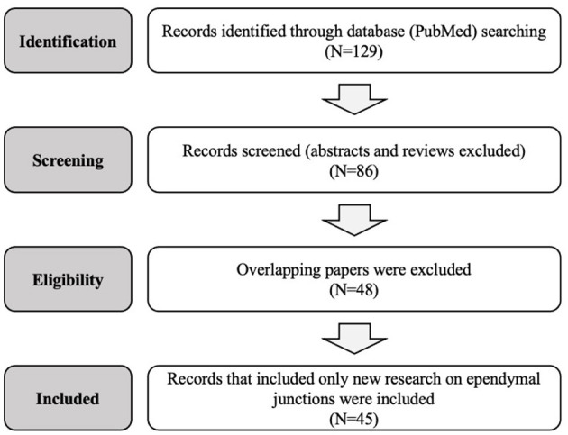
Methods: We systematically searched and reviewed original research articles treating ependymal intercellular junctions on PubMed. Reviews, opinion papers, and abstracts were excluded. Research conducted on tissue samples, cell lines, CSF, and animal models was considered.
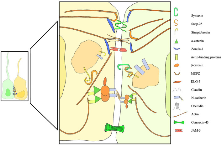
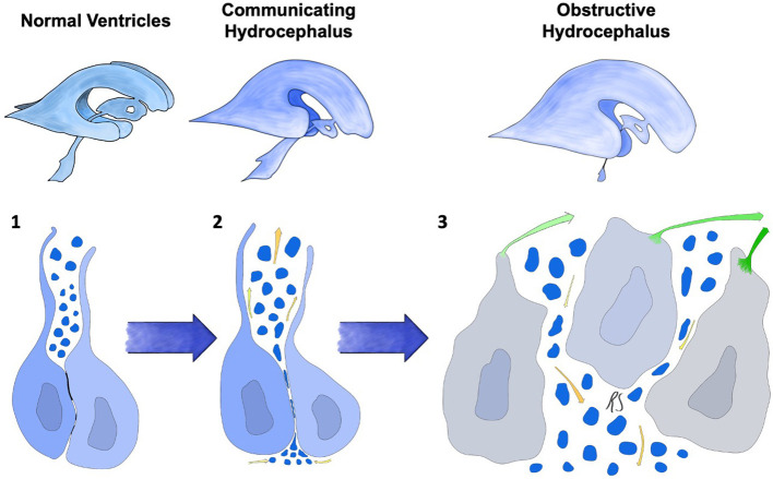
Results: A total of 45 novel articles treating tight, adherens and gap junctions of the ependyma were included in our review, spanning from 1960 to 2022. The findings of this review point toward a central and not yet fully characterized role of the ependymal lining ultrastructure in fluid flow interactions in the brain. In particular, tight junctions circumferentially line the apical equator of ependymal cells, changing between embryonal and adult life in several rodent models, shaping fluid and solute transit in this location. Further, adherens and gap junctions appear to have a pivotal role in several forms of congenital hydrocephalus.
Conclusions: These findings may provide an opportunity for medical management of CSF disorders, potentially allowing for tuning of CSF secretion and absorption. Beyond hydrocephalus, stroke, trauma, this information has relevance for metabolite clearance and drug delivery, with potential to affect many patients with a variety of neurological disorders. This critical look at intercellular junctions in ependyma and the surrounding interstitial spaces is meant to inspire future research on a central and rather unknown component of the CSF-brain interface.
Keywords: adherens junctions; ependyma; gap junctions; hydrocephalus; tight junctions.
Copyright © 2023 Serra and Simard.
Conflict of interest statement
The authors declare that the research was conducted in the absence of any commercial or financial relationships that could be construed as a potential conflict of interest.
Figures
Similar articles
-
Phorbol ester induced changes in tight and adherens junctions in the choroid plexus epithelium and in the ependyma.Brain Res. 2000 Jan 31;854(1-2):197-206. doi: 10.1016/s0006-8993(99)02355-0. Brain Res. 2000. PMID: 10784122
-
Disruption of CDH2/N-cadherin-based adherens junctions leads to apoptosis of ependymal cells and denudation of brain ventricular walls.J Neuropathol Exp Neurol. 2013 Sep;72(9):846-60. doi: 10.1097/NEN.0b013e3182a2d5fe. J Neuropathol Exp Neurol. 2013. PMID: 23965744
-
The mouse Jhy gene regulates ependymal cell differentiation and ciliogenesis.PLoS One. 2017 Dec 6;12(12):e0184957. doi: 10.1371/journal.pone.0184957. eCollection 2017. PLoS One. 2017. PMID: 29211732 Free PMC article.
-
Structure and function of the ependymal barrier and diseases associated with ependyma disruption.Tissue Barriers. 2014 Mar 19;2:e28426. doi: 10.4161/tisb.28426. eCollection 2014. Tissue Barriers. 2014. PMID: 25045600 Free PMC article. Review.
-
The biological significance of brain barrier mechanisms: help or hindrance in drug delivery to the central nervous system?F1000Res. 2016 Mar 10;5:F1000 Faculty Rev-313. doi: 10.12688/f1000research.7378.1. eCollection 2016. F1000Res. 2016. PMID: 26998242 Free PMC article. Review.
Cited by
-
Cellular junction dynamics and Alzheimer's disease: a comprehensive review.Mol Biol Rep. 2024 Feb 1;51(1):273. doi: 10.1007/s11033-024-09242-w. Mol Biol Rep. 2024. PMID: 38302794 Review.
-
Junctional adhesion molecule-C: A multifunctional mediator of cell adhesion.Cell Mol Life Sci. 2025 Aug 13;82(1):312. doi: 10.1007/s00018-025-05829-z. Cell Mol Life Sci. 2025. PMID: 40801950 Free PMC article. Review.
-
Multiciliated ependymal cells: an update on biology and pathology in the adult brain.Acta Neuropathol. 2024 Sep 10;148(1):39. doi: 10.1007/s00401-024-02784-0. Acta Neuropathol. 2024. PMID: 39254862 Review.
-
Hydrocephalus in Connection to Genetic Mutation in Cranial Neural Crest Cells.Orthod Craniofac Res. 2025 May 13:10.1111/ocr.12942. doi: 10.1111/ocr.12942. Online ahead of print. Orthod Craniofac Res. 2025. PMID: 40357558 Review.
-
The periaxonal space as a conduit for cerebrospinal fluid flow to peripheral organs.Proc Natl Acad Sci U S A. 2024 Nov 5;121(45):e2400024121. doi: 10.1073/pnas.2400024121. Epub 2024 Nov 1. Proc Natl Acad Sci U S A. 2024. PMID: 39485799 Free PMC article.
References
-
- Dandy WE, Blackfan KD. Internal hydrocephalus: second paper. Am J Dis Child. (1917) 14:424–43. 10.1001/archpedi.1917.01910120029002 - DOI
Publication types
LinkOut - more resources
Full Text Sources
Miscellaneous