. 2023 Apr 4;107(1):22.
doi: 10.5334/jbsr.3021. eCollection 2023.

CT-Angiographic Aspects of Pulmonary Embolism on SARS COV-2

Affiliations

CT-Angiographic Aspects of Pulmonary Embolism on SARS COV-2

Bénilde Marie-Ange Tiemtore-Kambou et al. J Belg Soc Radiol. .

Erratum in

  • Correction: CT-Angiographic Aspects of Pulmonary Embolism on SARS COV-2.
    Tiemtoré-Kambou BM, Ouédraogo A, Dao SBA, Sieba IFN, Koama A, Traore IS, Napon S, Diendéré ÉA, Ouédraogo W, Sankara HD, Cisse R. Tiemtoré-Kambou BM, et al. J Belg Soc Radiol. 2023 May 12;107(1):39. doi: 10.5334/jbsr.3202. eCollection 2023. J Belg Soc Radiol. 2023. PMID: 37188082 Free PMC article.

Abstract

Objectives: To study pulmonary embolism during COVID-19 pneumonia.

Patients and methods: This was a one-year retrospective and descriptive study of all patients from three imaging sites with SARS-CoV2 infection.

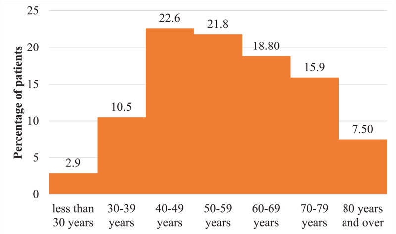
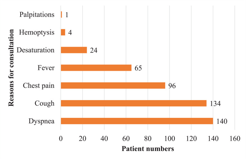
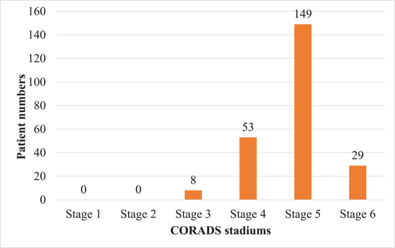
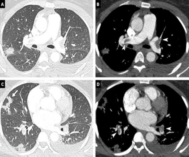
Results: Two hundred and thirty-nine patients were included. The prevalence of pulmonary embolism was 18.4%. The average age was 55 years old. The sex ratio was 1.65. Dyspnea (58.6%), cough (56.1%), and chest pain (40.2%) were the most common reasons for consultation. In 151 patients (63.2%), chest computed tomography (CT) angiography was performed without checking level of D-dimer. The level of D-dimers was elevated in 47.8%. Grade 5 of CO-RADS accounted for 62.3%. In 70.5% of cases, the pulmonary embolism was bilateral with subsegmental involvement in 47.7%.Condensation in 'ground glass' with 'crazy paving' were the predominant typical parenchymal lesions with a frequency of 93.7% and 59.4%. In univariate analysis, D-dimers were significantly associated with the occurrence of pulmonary embolism (p < 0.001). Male sex was associated with a non-significantly higher Risk of having a pulmonary embolism (1.18 95% CI: 0.61-2.31, p = 0.622). The critical level increased the risk of pulmonary embolism in a non-significant way. Only the high level of D-dimers was and this, in a significant way.

Conclusion: Pulmonary embolism was increased in the context of SARS-CoV2. The chest CT-angiography associated with the dosage of D-dimers constitutes a good diagnostic arsenal.

Keywords: COVID-19; D-dimers; chest CT angiography; pulmonary embolism.


Conflict of interest statement

The authors have no competing interests to declare.

Figures

Distribution of patients by age group
Figure 1
Distribution of patients by age group.
Distribution of patients by reason for consultation
Figure 2
Distribution of patients by reason for consultation.
Distribution of patients according to the CO-RADS classification
Figure 3
Distribution of patients according to the CO-RADS classification.
Chest CT-angiography of a 38-year-old patient with no pathological history with a positive PCR for COVID-19 pneumonia. Parenchymal window (A, C) shows peripheral foci of condensation. Mediastinal window (B, D) finds a bilateral proximal pulmonary embolism (endoluminal defects at the bilateral lobar level) as well as a left pleural effusion
Figure 4
Chest CT-angiography of a 38-year-old patient with no pathological history with a positive PCR for COVID-19 pneumonia. Parenchymal window (A, C) shows peripheral foci of condensation. Mediastinal window (B, D) finds a bilateral proximal pulmonary embolism (endoluminal defects at the bilateral lobar level) as well as a left pleural effusion.
Thoracic CT angiography of a 42-year-old patient with positive PCR for COVID-19. Parenchymal window in the axial section (A) without injection shows peripheral bilateral ‘ground glass’ areas (arrow). The mediastinal window (B) in coronal reconstruction (MIP), reveals several bilateral endoluminal defects involving the lobar, segmental, and sub-segmental branches of the pulmonary artery (arrow)
Figure 5
Thoracic CT angiography of a 42-year-old patient with positive PCR for COVID-19. Parenchymal window in the axial section (A) without injection shows peripheral bilateral ‘ground glass’ areas (arrow). The mediastinal window (B) in coronal reconstruction (MIP), reveals several bilateral endoluminal defects involving the lobar, segmental, and sub-segmental branches of the pulmonary artery (arrow).

References

    1. Revel MP, Parkar AP, Prosch H, et al. European Society of Radiology (ESR) and European Society of Thoracic Imaging (ESTI). COVID-19 patients and the radiology department – advice from the European Society of Radiology (ESR) and the European Society for Thoracic Imaging (ESTI). EUR Radiol. 2020; 30: 4903–4909. DOI: 10.1007/s00330-020-06865-y - DOI - PMC - PubMed
    1. Bompard F, Monnier H, Saab I, et al. Pulmonary embolism in patients with COVID-19 pneumonia. EUR Breathe J. 2020; 56: 2001365. DOI: 10.1183/13993003.01365-2020 - DOI - PMC - PubMed
    1. Guzik TJ, Mohiddin SA, Dimarco A, et al. COVID-19 and the cardiovascular system: Implications for risk assessment, diagnosis, and treatment options. Cardiovasc Res; 2020. DOI: 10.1093/cvr/cvaa106 - DOI - PMC - PubMed
    1. Prokop M, van Everdingen W, van Rees Vellinga T, et al. CO-RADS: A categorical CT assessment scheme for patients suspected of having COVID-19—Definition and evaluation. Radiology. 2020; 296: E97–104. DOI: 10.1148/radiol.2020201473 - DOI - PMC - PubMed
    1. http://ebulletin.radiologie.fr/covid19.

LinkOut - more resources