Review
. 2023 Mar;4(1):011315.
doi: 10.1063/5.0127713. Epub 2023 Mar 30.

Emerging methods to model cardiac ion channel and myocyte electrophysiology

Affiliations
Review

Emerging methods to model cardiac ion channel and myocyte electrophysiology

Jonathan D Moreno et al. Biophys Rev (Melville). 2023 Mar.

Abstract

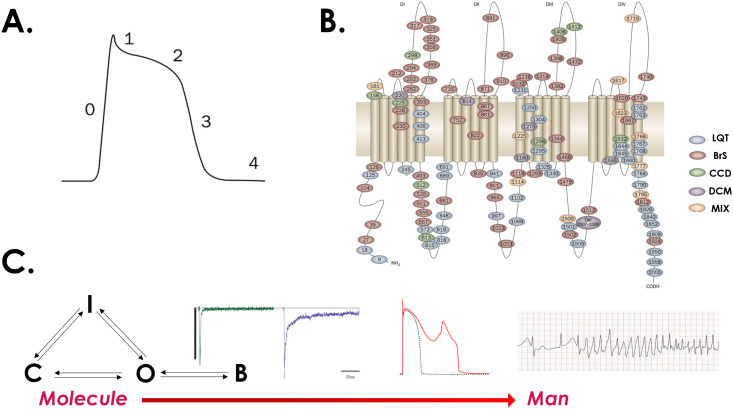
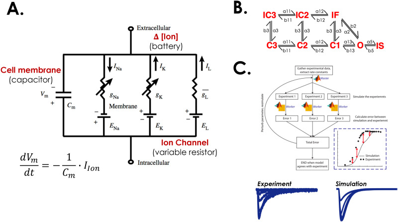
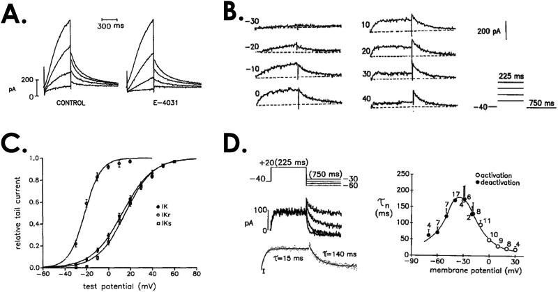
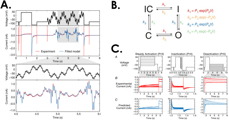
In the field of cardiac electrophysiology, modeling has played a central role for many decades. However, even though the effort is well-established, it has recently seen a rapid and sustained evolution in the complexity and predictive power of the models being created. In particular, new approaches to modeling have allowed the tracking of parallel and interconnected processes that span from the nanometers and femtoseconds that determine ion channel gating to the centimeters and minutes needed to describe an arrhythmia. The connection between scales has brought unprecedented insight into cardiac arrhythmia mechanisms and drug therapies. This review focuses on the generation of these models from first principles, generation of detailed models to describe ion channel kinetics, algorithms to create and numerically solve kinetic models, and new approaches toward data gathering that parameterize these models. While we focus on application of these models for cardiac arrhythmia, these concepts are widely applicable to model the physiology and pathophysiology of any excitable cell.


Conflict of interest statement

The authors have no conflicts to disclose.

Figures

FIG. 1.
FIG. 1.
The generation of the cardiac action potential in both health and disease. (a) The cardiac ventricular action potential is divided into five phases, with the rapid influx of Na+ into the cell leading to the fast upstroke of the AP (phase 0). (b) Numerous diseases have been linked to the cardiac Na+ channel including the long QT3, Brugada, and dilated cardiomyopathy. (c) In the prototypical LQT3 mutation, some channels fail to inactivate and enter a bursting regime (denoted B). This leads to a small, sustained inward Na+ current (purple trace). At the single cell level, this manifests as AP prolongation (red waveform, depicting an early afterdepolarization). At the organ level, this can lead to the deadly rhythm disturbance known as polymorphic ventricular tachycardia. Adapted with permission from Ruan et al., Nat. Rev. Cardiol. 6, 337–348. 2009, Copyright 2009 Nature Publishing.
FIG. 2.
FIG. 2.
Creation and numerical optimization of quantitative descriptions of ion channels. (a) A simple circuit diagram representing the cell membrane (capacitor), and three ion channels (gNa, gK, and gL) representing variable resistors. (b) A prototypical eight-state Na+ channel Markov model with three closed states, one open state, two closed inactivated states and a fast and slow inactivated state arising from the open state. (c) To fit the rate constants depicted in (b), numerical optimization techniques are employed to simulate an electrophysiologic experiment. The simulated results are compared to the experiment, and the rate constants are adjusted to minimize the objective function (the error between simulation and experiment. Adapted with permission from Y. Rudy and J. R. Silva, Q. Rev. Biophys. 39(1), 57–116 (2006). Copyright 2006 by Cambridge University Press. In addition, Adapted with permission from Moreno et al., PLoS One 11(3), e0150761 (2016). Copyright 2016 Authors, licensed under a Create Commons Attribution (CC BY) License.
FIG. 3.
FIG. 3.
Traditional protocols that are used to characterize ion channel time- and voltage-dependent kinetics. (a) K+ currents that arise from a series of steps to depolarized potentials from a guinea pig ventricular myocyte. A component of the overall current (left) is blocked by the molecule E-4031 (right). (b) Subtraction of the E-4031 blocked current from control yields the difference current. In this case, the rapid component of the delayed rectifier K+ channel, IKr. Shown is the response to steps at different potentials according to the protocol shown at the bottom right. (c) The relationship between the steady state conductance and the membrane potential is calculated by dividing the peak current magnitude by the driving force, e.g., (Vm − EK) where Vm is the membrane potential and EK is the reversal potential for K+, providing n for the model. (d) The time constant τn is found by fitting the activation at depolarized potentials as in (b), and hyperpolarizing potentials after activation according to the protocol at the top left of the panel which steps to different negative potentials. Adapted with permission from Sanguinetti et al., J. Gen. Physiol. 96(1), 195–215 (1990). Copyright 1990 by Rockefeller University Press.
FIG. 4.
FIG. 4.
Sinusoidal waveforms can be used to parameterize Markov models. (a) The waveform at the top is used to evoke the current below and this is fit to parameters for a four-state Markov model. (b) The Markov model that was fit to the parameters includes four states with forward and backward rates between each of the states. The rates are described by exponential equations. (c) A test of the model evaluated whether it could predict ionic currents from traditional square pulse protocols, and it was able to do so successfully. Adapted with permission from Beattie et al., J. Physiol. 596(10), 1813–1828 (2018). Copyright 2018 by The Physiological Society.

Similar articles

References

    1. Hodgkin A. L. and Huxley A. F., “ A quantitative description of membrane current and its application to conduction and excitation in nerve,” J. Physiol. 117(4), 500–544 (1952).10.1113/jphysiol.1952.sp004764 - DOI - PMC - PubMed
    1. Rudy Y. and Silva J. R., “ Computational biology in the study of cardiac ion channels and cell electrophysiology,” Q. Rev. Biophys. 39(1), 57–116 (2006).10.1017/S0033583506004227 - DOI - PMC - PubMed
    1. Ruan Y., Liu N., and Priori S. G., “ Sodium channel mutations and arrhythmias,” Nat. Rev. Cardiol. 6, 337–348 (2009).10.1038/nrcardio.2009.44 - DOI - PubMed
    1. Luo C. H. and Rudy Y., “ A dynamic model of the cardiac ventricular action potential. I. Simulations of ionic currents and concentration changes,” Circ. Res. 74(6), 1071–1096 (1994).10.1161/01.RES.74.6.1071 - DOI - PubMed
    1. Campbell D. L. et al., “ Reversal potential of the calcium current in bull-frog atrial myocytes,” J. Physiol. 403, 267–286 (1988).10.1113/jphysiol.1988.sp017249 - DOI - PMC - PubMed

LinkOut - more resources