

Development of stromal differentiation patterns in heterotypical models of artificial corneas generated by tissue engineering
- PMID: 37034263
- PMCID: PMC10076743
- DOI: 10.3389/fbioe.2023.1124995
Development of stromal differentiation patterns in heterotypical models of artificial corneas generated by tissue engineering
Abstract
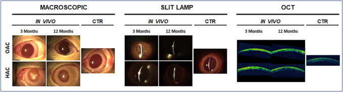
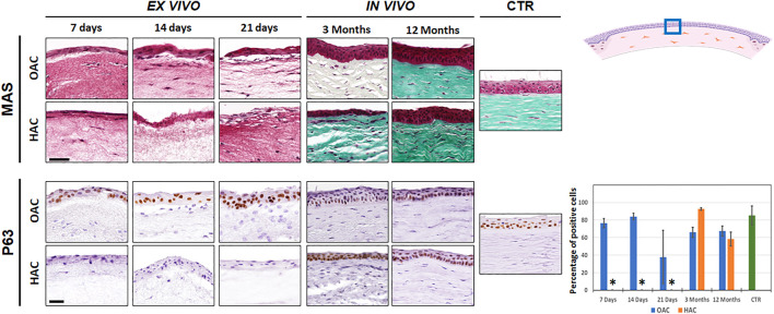
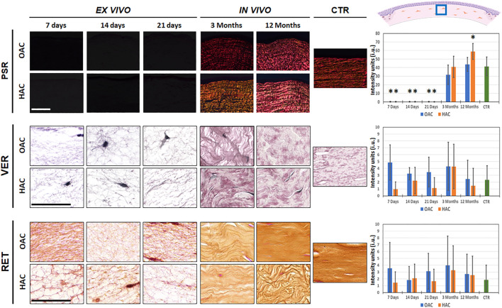
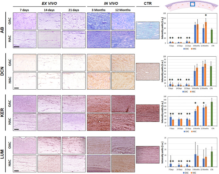
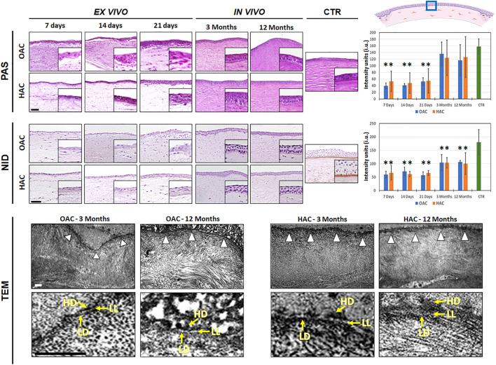
Purpose: We carried out a histological characterization analysis of the stromal layer of human heterotypic cornea substitutes generated with extra-corneal cells to determine their putative usefulness in tissue engineering. Methods: Human bioartificial corneas were generated using nanostructured fibrin-agarose biomaterials with corneal stromal cells immersed within. To generate heterotypical corneas, umbilical cord Wharton's jelly stem cells (HWJSC) were cultured on the surface of the stromal substitutes to obtain an epithelial-like layer. These bioartificial corneas were compared with control native human corneas and with orthotypical corneas generated with human corneal epithelial cells on top of the stromal substitute. Both the corneal stroma and the basement membrane were analyzed using histological, histochemical and immunohistochemical methods in samples kept in culture and grafted in vivo for 12 months in the rabbit cornea. Results: Our results showed that the stroma of the bioartificial corneas kept ex vivo showed very low levels of fibrillar and non-fibrillar components of the tissue extracellular matrix. However, in vivo implantation resulted in a significant increase of the contents of collagen, proteoglycans, decorin, keratocan and lumican in the corneal stroma, showing higher levels of maturation and spatial organization of these components. Heterotypical corneas grafted in vivo for 12 months showed significantly higher contents of collagen fibers, proteoglycans and keratocan. When the basement membrane was analyzed, we found that all corneas grafted in vivo showed intense PAS signal and higher contents of nidogen-1, although the levels found in human native corneas was not reached, and a rudimentary basement membrane was observed using transmission electron microscopy. At the epithelial level, HWJSC used to generate an epithelial-like layer in ex vivo corneas were mostly negative for p63, whereas orthotypical corneas and heterotypical corneas grafted in vivo were positive. Conclusion: These results support the possibility of generating bioengineered artificial corneas using non-corneal HWJSC. Although heterotypical corneas were not completely biomimetic to the native human corneas, especially ex vivo, in vivo grafted corneas demonstrated to be highly biocompatible, and the animal cornea became properly differentiated at the stroma and basement membrane compartments. These findings open the door to the future clinical use of these bioartificial corneas.
Keywords: Wharton’s jelly mesenchymal stem cells; basement membrane; cornea; stroma; tissue engineering.
Copyright © 2023 Blanco-Elices, Morales-Álvarez, Chato-Astrain, González-Gallardo, Ávila-Fernández, Campos, Carmona, Martín-Piedra, Garzón and Alaminos.
Conflict of interest statement
The authors declare that the research was conducted in the absence of any commercial or financial relationships that could be construed as a potential conflict of interest.
Figures
Similar articles
-
Histological, histochemical, and immunohistochemical characterization of NANOULCOR nanostructured fibrin-agarose human cornea substitutes generated by tissue engineering.BMC Med. 2024 Nov 13;22(1):531. doi: 10.1186/s12916-024-03759-4. BMC Med. 2024. PMID: 39538248 Free PMC article.
-
Long-Term in vivo Evaluation of Orthotypical and Heterotypical Bioengineered Human Corneas.Front Bioeng Biotechnol. 2020 Jun 19;8:681. doi: 10.3389/fbioe.2020.00681. eCollection 2020. Front Bioeng Biotechnol. 2020. PMID: 32671048 Free PMC article.
-
Generation of a biomimetic human artificial cornea model using Wharton's jelly mesenchymal stem cells.Invest Ophthalmol Vis Sci. 2014 Jun 6;55(7):4073-83. doi: 10.1167/iovs.14-14304. Invest Ophthalmol Vis Sci. 2014. PMID: 24906855
-
The corneal fibrosis response to epithelial-stromal injury.Exp Eye Res. 2016 Jan;142:110-8. doi: 10.1016/j.exer.2014.09.012. Exp Eye Res. 2016. PMID: 26675407 Free PMC article. Review.
-
Corneal regeneration: A review of stromal replacements.Acta Biomater. 2018 Mar 15;69:31-41. doi: 10.1016/j.actbio.2018.01.023. Epub 2018 Feb 1. Acta Biomater. 2018. PMID: 29374600 Review.
Cited by
-
A novel 3D biofabrication strategy to improve cell proliferation and differentiation of human Wharton's jelly mesenchymal stromal cells for cell therapy and tissue engineering.Front Bioeng Biotechnol. 2023 Aug 10;11:1235161. doi: 10.3389/fbioe.2023.1235161. eCollection 2023. Front Bioeng Biotechnol. 2023. PMID: 37636000 Free PMC article.
-
Spatiotemporal characterization of extracellular matrix maturation in human artificial stromal-epithelial tissue substitutes.BMC Biol. 2024 Nov 18;22(1):263. doi: 10.1186/s12915-024-02065-y. BMC Biol. 2024. PMID: 39558321 Free PMC article.
-
A Novel In Vitro Pathological Model for Studying Neural Invasion in Non-Melanoma Skin Cancer.Gels. 2024 Apr 8;10(4):252. doi: 10.3390/gels10040252. Gels. 2024. PMID: 38667671 Free PMC article.
-
Histological, histochemical, and immunohistochemical characterization of NANOULCOR nanostructured fibrin-agarose human cornea substitutes generated by tissue engineering.BMC Med. 2024 Nov 13;22(1):531. doi: 10.1186/s12916-024-03759-4. BMC Med. 2024. PMID: 39538248 Free PMC article.
References
-
- Alfonso-Rodríguez C. A., González-Andrades E., Jaimes-Parra B. D., Fernández-Valadés R., Campos A., Sánchez-Quevedo M. C., et al. (2015). Ex vivo and in vivo modulatory effects of umbilical cord Wharton’s jelly stem cells on human oral mucosa stroma substitutes. Histol. Histopathol. 30, 1321–1332. 10.14670/HH-11-628 - DOI - PubMed
-
- Borys-Wójcik S., Brązert M., Jankowski M., Ożegowska K., Chermuła B., Piotrowska-Kempisty H., et al. (2019). Human Wharton’s jelly mesenchymal stem cells: Properties, isolation and clinical applications. J. Biol. Regul. Homeost. Agents 33, 119–123. - PubMed
LinkOut - more resources
Full Text Sources