Case Reports
. 2023 Mar 24:10:1123789.
doi: 10.3389/fcvm.2023.1123789. eCollection 2023.

Multimodality imaging of a cardiac paraganglioma: A case report

Affiliations
Case Reports

Multimodality imaging of a cardiac paraganglioma: A case report

Bruna Punzo et al. Front Cardiovasc Med. .

Abstract

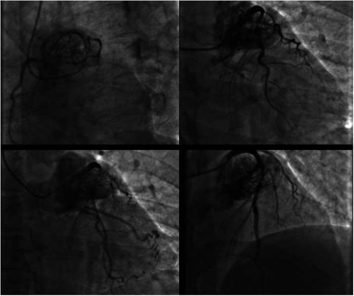
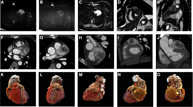
Cardiac paragangliomas (PGLs) are rare extra-adrenal tumors that arise from chromaffin cells of the sympathetic ganglia. PGLs are often diagnosed incidentally, in the absence of symptoms, or with symptoms related to cardiovascular dysfunction. Cardiac computed tomography (CCT) and cardiac magnetic resonance (CMR) can be used to accurately determine the lesion morphology and position as well as providing detailed tissue characterization. A multimodal imaging approach, not yet standardized, could be useful either in diagnosis and monitoring or in treatment planning. In the case reported here, CCT and CMR were performed to define lesion anatomy, and a reconstruction was generated using cinematic rendering (CR) to characterize the PGL angioarchitecture.

Keywords: CCT; CMR; cardiac paraganglioma; case report; neuroendocrine tumor.


Conflict of interest statement

The authors declare that the research was conducted in the absence of any commercial or financial relationships that could be construed as a potential conflict of interest.

Figures

Figure 1
Figure 1
Coronary angiogram in different oblique projections showing a large cardiac mass encasing the left coronary artery.
Figure 2
Figure 2
CMR acquisition (A–E): DWI b 0 and b 1,000 (A,B) and ADC map (C) showing high cellularity of the lesion; balanced gradient echo sequence in short axis and two chambers (D,E), showing morphology and extension of a PGL. CCT acquisition (F–J): axial (F,G) and oblique planes highlighting the morphology, position, and contrast of the PGL. CR reconstructions in different planes (K–O).

References

    1. Nemeth A, Schlensak C, Popov A. Extended resection of a cardiac paraganglioma – a rare neuroendocrine manifestation of the heart. J Card Surg. (2020) 35(3):700–2. 10.1111/jocs.14440 - DOI - PubMed
    1. Yadav PK, Baquero GA, Malysz J, Kelleman J, Gilchrist IC. Cardiac paraganglioma. Circ Cardiovasc Interv. (2014) 7(6):851–6. 10.1161/CIRCINTERVENTIONS.114.001856 - DOI - PubMed
    1. Bhojwani N, Huang J, Garg V, Yang M, Oliveira GH, Rajiah P. Utility of 18F-fluorodeoxyglucose positron emission tomography/magnetic resonance imaging in the diagnosis of cardiac paraganglioma. Indian J Nucl Med. (2017) 32(4):380–2. 10.4103/ijnm.IJNM_93_17 - DOI - PMC - PubMed
    1. Eid M, De Cecco CN, Nance JW, Caruso D, Albrecht MH, Spandorfer AJ, et al. Cinematic rendering in CT: a novel, lifelike 3D visualization technique. Am J Roentgenol. (2017) 209(2):370–9. 10.2214/AJR.17.17850 - DOI - PubMed
    1. Saththasivam P, Herrera E, Jabbari OA, Reardon M, Sheinbaum R. Cardiac paraganglioma resection with ensuing left main coronary artery compromise. J Cardiothorac Vasc Anesth. (2017) 31(1):236–9. 10.1053/j.jvca.2016.05.048 - DOI - PubMed

Publication types