[Preprint]. 2023 Mar 31:2023.03.28.534602.
doi: 10.1101/2023.03.28.534602.

Deep repertoire mining uncovers ultra-broad coronavirus neutralizing antibodies targeting multiple spike epitopes

Affiliations

Deep repertoire mining uncovers ultra-broad coronavirus neutralizing antibodies targeting multiple spike epitopes

Jonathan Hurtado et al. bioRxiv. .

Update in

Abstract

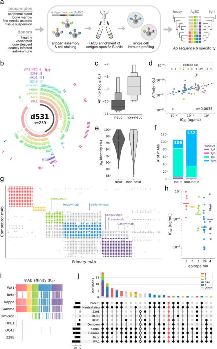
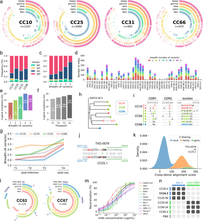
Development of vaccines and therapeutics that are broadly effective against known and emergent coronaviruses is an urgent priority. Current strategies for developing pan-coronavirus countermeasures have largely focused on the receptor binding domain (RBD) and S2 regions of the coronavirus Spike protein; it has been unclear whether the N-terminal domain (NTD) is a viable target for universal vaccines and broadly neutralizing antibodies (Abs). Additionally, many RBD-targeting Abs have proven susceptible to viral escape. We screened the circulating B cell repertoires of COVID-19 survivors and vaccinees using multiplexed panels of uniquely barcoded antigens in a high-throughput single cell workflow to isolate over 9,000 SARS-CoV-2-specific monoclonal Abs (mAbs), providing an expansive view of the SARS-CoV-2-specific Ab repertoire. We observed many instances of clonal coalescence between individuals, suggesting that Ab responses frequently converge independently on similar genetic solutions. Among the recovered antibodies was TXG-0078, a public neutralizing mAb that binds the NTD supersite region of the coronavirus Spike protein and recognizes a diverse collection of alpha- and beta-coronaviruses. TXG-0078 achieves its exceptional binding breadth while utilizing the same VH1-24 variable gene signature and heavy chain-dominant binding pattern seen in other NTD supersite-specific neutralizing Abs with much narrower specificity. We also report the discovery of CC24.2, a pan-sarbecovirus neutralizing mAb that targets a novel RBD epitope and shows similar neutralization potency against all tested SARS-CoV-2 variants, including BQ.1.1 and XBB.1.5. A cocktail of TXG-0078 and CC24.2 provides protection against in vivo challenge with SARS-CoV-2, suggesting potential future use in variant-resistant therapeutic Ab cocktails and as templates for pan-coronavirus vaccine design.


Conflict of interest statement

Competing interests D.B.J., B.A.A., M.J.T.S., and W.J.M. were employees and shareholders of 10x Genomics, Inc. at the time this work was performed. D.B.J., B.A.A., M.J.T.S., and W.J.M. are inventors on patents in relation to algorithms, therapeutic candidates, and other components of this work.

Figures

Figure 1.
Figure 1.. Discovery of an ultra-broad neutralizing antibody targeting the NTD.
(a) Overview of the multiplexed antigen screening method. (b) Binding specificities of recombinantly expressed mAbs from Donor 531 (d531). Each arc segment corresponds to a single mAb, and the ring corresponding to each Spike antigen is colored if bound by the mAb. The centermost band is colored dark gray if the mAb neutralizes SARS-CoV-2 and light gray if non-neutralizing. (c) Letter-value plot of mAb affinity for WA1 Spike. The center line on each box represents the median. (d) Correlation between binding affinity (KD) and neutralization (IC50) for the WA1 strain of SARS-CoV-2. The linear regression line is shown, as well as the p-value indicating a significant positive slope. (e) Violin plot of heavy chain Variable (VH) germline gene identity. 100% identity indicates no somatic mutation. (f) Isotype distribution of WA1 nAbs and non-nAbs. The total number of mAbs in each category (neut and non-neut) is shown at the top of the respective bar. (g) Epitope binning by competitive binding inhibition. For each mAb pair, a darkly filled circle indicates strong competition, a lightly filled circle indicates weak competition, and no circle indicates no competition. Points are colored according to their respective epitope bin. (h) Neutralization potency against SARS-CoV-2 of nAbs in each epitope bin. Bins are colored as in (g). (i) Heatmap of mAb binding affinity (KD) for SARS-CoV-2 VoCs and seasonal CoV S-proteins. Colors correspond to epitope bins and are the same as in (g). Color intensity corresponds to binding affinity, with high affinity colored most intensely. (j) Upset plot showing cross-reactivity binding and neutralization patterns among epitope binned mAbs. The broadest cross-reactivity group, which bind all tested CoV S-proteins but do not neutralize, are indicated by unfilled dots. TXG-0078, which comprises its own cross-reactivity pattern, is highlighted in pink.
Figure 2.
Figure 2.. Quantifying the development of mAb breadth of binding using antigen barcodes.
(a) Ring plots showing the antigen barcode classifications of 6,305 SARS-CoV-2-specific mAbs from participants CC10, CC25, CC31 and CC66. Individual mAbs are represented by radial vectors, with each antigen ring (WA1: blue, alpha: green, beta: yellow, gamma: orange, kappa: magenta) colored if the given B cell was classified as positive for the respective antigen. Antibodies are sorted by breadth, with breadth decreasing in the clockwise direction. (b) Isotype distribution of SARS-CoV-2-specific mAbs for each donor. (c) Isotype distribution of mAbs, grouped by cross-variant breadth. (d) Heavy chain Variable (VH) gene use of SARS-CoV-2-speicific mAbs, grouped by cross-variant breadth. (e) Number of VH nucleotide mutations in SARS-CoV-2-specific mAbs, grouped by breadth. (f) Number of VH nucleotide mutations in SARS-CoV-2-specific mAbs, grouped by timepoint (T1, T2 and T3 are post-infection but pre-vaccination; T4 is post-infection and post-vaccination). (g) Median cross-variant breadth of all mAbs isolated at each timepoint, grouped by donor. The dashed black line represents the median of all mAbs from participants. (h) Phylogenetic representation of a single public SARS-CoV-2-specific clonotype. All mAbs in the clonotype use the IgG1 isotype except for a single IgA1 clone (indicated by text and an unfilled marker). (i) Complementarity determining region (CDR) alignments of sequences in the public clonotype shown in (h). Mutations (CDR1 and 2) or untemplated residues (Junction) shared between multiple participants are highlighted in color (CC10: magenta, CC25: green, CC66: blue). The lone IgA1 mAb is indicated by an unfilled marker. (j) Junction alignments of a second public clonotype, comprising TXG-0078 (from donor 531) and CC25.1 (from donor CC25). Untemplated positions (N- and P-addition) are indicated in bold, and identical residues in untemplated regions are highlighted in red. (k) Distribution of cross-donor alignment scores obtained by iterative pairwise alignment of heavy chain amino acid sequences. Only sequences recovered from different doors were compared. Distributions resulting from comparison of sequences encoding the same V gene (green), V genes from the same family (orange) or V genes from different families (blue) are shown. The alignment score between TXG-0078 and CC25.1 is highlighted. (l) Ring plots showing antigen barcode classifications of two additional participants (CC62 and CC67). Antigen rings are colored if the respective mAb was classified as antigen positive (WA1: magenta, beta: yellow, kappa: green, SARS-CoV: blue, YN02 RBD: purple). (m) Neutralization of SARS-CoV (red) and several SARS-CoV-2 variants by mAb CC24.2. (n) Epitope binning of mAb CC24.2 with several RBD-specific mAbs. Two competitor mAbs (CC25.4 and CC25.56) bind the Site 5 epitope, the other competitor mAbs (CC25.54, CC84.24 and CC84.2) bind other RBD epitopes. Binding inhibition (dark gray) or lack of binding inhibition (light gray) is shown, with phosphate buffered saline (PBS) used as a negative control.
Figure 3.
Figure 3.. Structural definition of the broad, NTD-supersite antibody TXG-0078.
(a) Representative 2D classes obtained by cryo-EM analysis of TXG-0078 Fab in complex with SARS-CoV-2 spike (b) Two cryo-EM reconstructions of the TXG-0078-spike complex at 3.14 Å (Rosy Brown/Grey) and 3.34 Å (Red) resolutions showing conformational flexibility of the Fab-NTD region (c) Cryo-EM reconstruction (3.9Å) of the NTD-Fab region obtained by local refinement. The spike NTD is colored grey and the Fab is colored rosy brown (d) The atomic model of TXG-0078 Fab Fv bound to NTD docked in the local refinement map. (e) The atomic model reveals that TXG-0078 targets the NTD supersite as has been described for other antibodies, though it is capable of weakly binding endemic human coronaviruses. The N3 and N5 loops comprise the epitope of TXG-0078; the CDRH3 of TXG-0078 reaches into a pocket in the N5 loop and the light chain is only minimally engaged in binding. Epitope and paratope residues are colored in light blue and red, respectively.
Figure 4.
Figure 4.. TXG-0078 protects transgenic ACE2 mice from live SARS-CoV-2 challenge.
(a) Mice were administered 300 μg of TXG-0078, CC24.2, a cocktail of both TXG-0078 and CC24.2, or a control antibody targeting Zika virus (ZIKV-1). Mean percent change in baseline weight is shown for each group. Shading indicates standard error. Significant differences between groups (p≤0.05) are indicated by asterisks in the color of each group displaying significantly different weight loss than the control (ZIKV-1) group. Significance was determined using a one-way ANalysis Of VAriance (ANOVA) with Dunnet’s multiple comparisons test. (b) Percent of baseline weight at day 5 is shown for all mice, separated by treatment group.

References

    1. Drosten C, Günther S, Preiser W, van der Werf S, Brodt H-R, Becker S, et al. Identification of a novel coronavirus in patients with severe acute respiratory syndrome. N Engl J Med. 2003;348: 1967–1976. - PubMed
    1. Pollack MP, Pringle C, Madoff LC, Memish ZA. Latest outbreak news from ProMED-mail: novel coronavirus -- Middle East. Int J Infect Dis. 2013;17: e143–4. - PMC - PubMed
    1. Adam D. The pandemic’s true death toll: millions more than official counts. In: Nature Publishing Group UK; [Internet]. 18 Jan 2022. [cited 31 May 2022]. doi:10.1038/d41586-022-00104-8 - DOI - PubMed
    1. Andrews N, Stowe J, Kirsebom F, Toffa S, Rickeard T, Gallagher E, et al. Covid-19 Vaccine Effectiveness against the Omicron (B.1.1.529) Variant. N Engl J Med. 2022;386: 1532–1546. - PMC - PubMed
    1. Sette A, Crotty S. Adaptive immunity to SARS-CoV-2 and COVID-19. Cell. 2021;184: 861–880. - PMC - PubMed

Publication types