[Preprint]. 2023 Mar 30:rs.3.rs-2698138.
doi: 10.21203/rs.3.rs-2698138/v1.

Astrocyte-neuron lactate shuttle plays a pivotal role in sensory-based neuroprotection in a rat model of permanent middle cerebral artery occlusion

Affiliations

Astrocyte-neuron lactate shuttle plays a pivotal role in sensory-based neuroprotection in a rat model of permanent middle cerebral artery occlusion

Mehwish Bhatti et al. Res Sq. .

Update in

Abstract

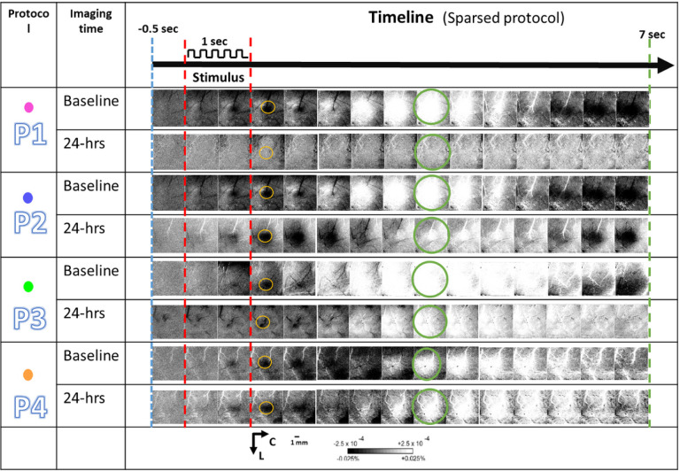
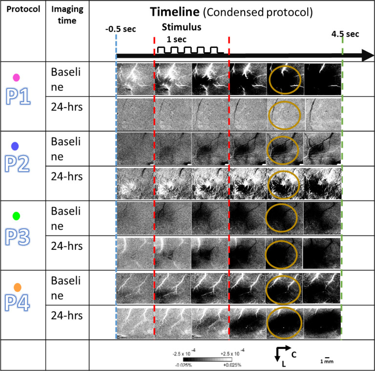
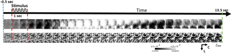
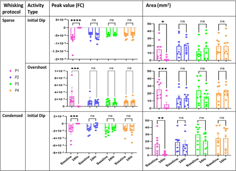
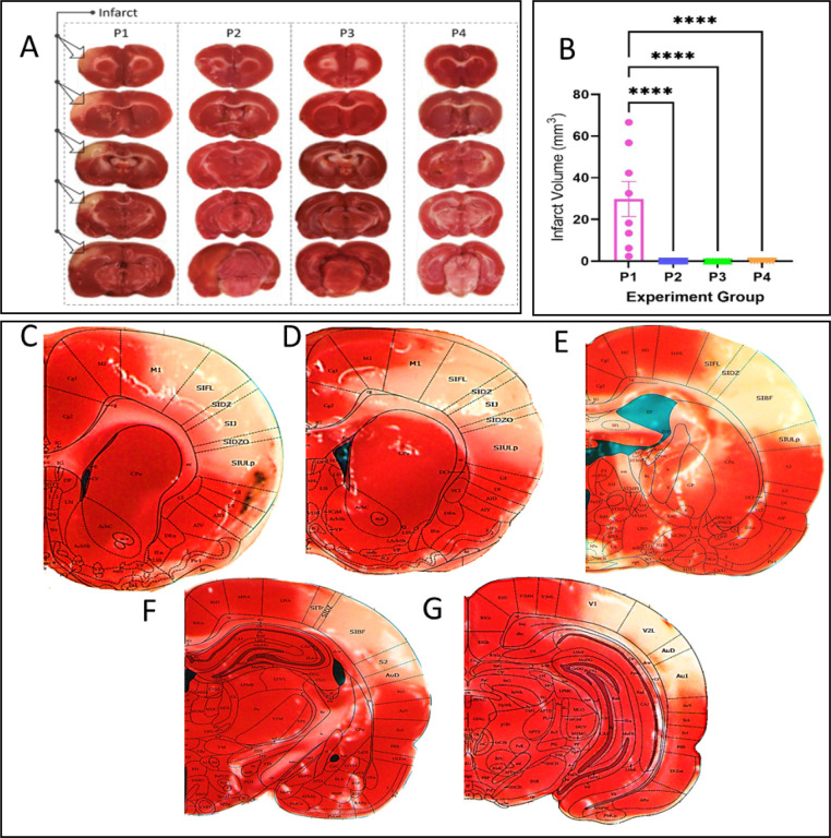
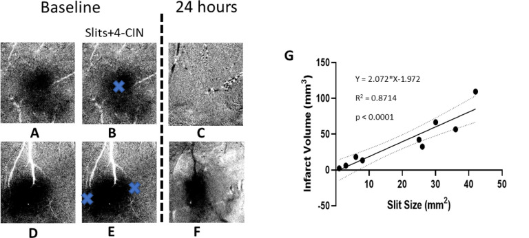
We have previously demonstrated protection from impending cortical stroke is achievable by sensory stimulation of the ischemic area in an adult rat model of permanent middle cerebral artery occlusion (pMCAo). We have further demonstrated that a major underpinning mechanism that is necessary for such protection is the system of collaterals among cerebral arteries that results in reperfusion of the MCA ischemic territory. However, since such collateral flow is weak, it may be necessary but not sufficient for protection and therefore we were seeking other complementary mechanisms that contribute to sensory-based protection. We hypothesized that astrocytes-to-neuron shuttle (ANLS) is another potential underpinning mechanism that could complement collateral flow in the protection process. Supporting our hypothesis, using functional imaging, pharmacological treatments, and postmortem histology, we show that ANLS has a pivotal role in sensory-based protection of cortex and therefor serves as the other supporting mechanism underpinning the protection process.


Conflict of interest statement

Competing interests (mandatory)

The author declares no competing interests.

Figures

Figure 1
Figure 1
Experimental design and surgical procedures. A) The detailed timeline of the experiment. B) Black and white arrowhead (top) show bregma and lambda respectively. The thick blue line shows the area thinned for ISOI. Small green area is thinned for pMCAo. C) Thinned area (approximate thickness range 24–32 um) prepared for ISOI; underlying cortex and vessels are clearly visible. D) The MCA-treatment, drug intervention details and the assigned color for each experiment group are tabulated. E) MCA after craniotomy and durotomy. Black arrowhead points to dorsal MCA. F) Two segments of thread are passed under MCA and loosely knotted G) Doubly knotted MCA. H) A petroleum jelly (Vaseline) well filled with saline/drug. I&J) tabulates parameters of sparse and condensed whisker stimulation protocols, respectively.
Figure 2
Figure 2
Aligned skull and dura slits. A) A small opening is made in the skull to lift part of it. The white streak is the reflection of overhead light. B) Blue arrows show lifted up skull. C) A small opening is made in dura with 30G bent needle hook. D&E) dura slit of desired size is made. Blue arrows show retraced dura after the slit at 120X magnification. F) The skull-slit halfway toward its original position, blue arrow. G) aligned dura-skull slits at 150X magnification bounded by blue dotted triangle.
Figure 3
Figure 3
A representative result for each experimental group following the sparse whisker stimulation protocol. The ISOI results at baseline (before pMCAo and drug diffusion) and at 24 hours (after pMCAo and drug diffusion) are shown for each experimental protocol (P1-P4). The red dotted lines denote the start and end of 1 sec 5hz-whisker stimulation denoted on top as a pulse train. The yellow and green circles show the specific frames which include the regions of interest used for quantification of initial dip and overshoot, respectively. Linear gray scale bar indicates intrinsic signal strength, C and L denotes caudal and lateral respectively. Each frame is ~7 mm × 7 mm. Black and white streaks are large surface blood vessels.
Figure 4
Figure 4
A representative result for each group for condensed whisker stimulation protocol. The ISOI results at baseline (before pMCAo and drug diffusion) and 24 hours after pMCAo and drug diffusion for each experimental protocol (P1-P4) are shown for comparison. The yellow circles show the specific frames which include the regions of interest used for quantification of initial dip. Bars and representative signs follow Figure 3.
Figure 5
Figure 5
ISOI-WFR of whisker C2 during sparse stimulation protocol. Top raw shows typical ISOI-WFR 3 phases response to stimulation during baseline, whereas the bottom raw shows the absence of the ISOI-WFR in presence of MCT inhibitor.
Figure 6
Figure 6
The spatial quantification of ISOI-WFR phases for the two stimulation paradigms. Top row: peak amplitude and area of the initial dip following application of the sparse whisker stimulation protocol as calculated at baseline and 24-hrs for each of the experimental groups P1-P4. Graphs show a significant difference only for group P1 at baseline and 24-hrs for both peak and area (****p<0.0001 and *p < 0.05). Middle row: peak amplitude and area of the overshoot calculated at baseline and 24-hrs following sparse protocol is shown for each of the experimental groups P1-P4 A significant difference is evident only for the P1 at baseline and at 24-hrs (***p<0.0021). Bottom row: peak amplitude and area of the initial dip following condensed whisker stimulation protocol as calculated at baseline and 24 hours for each of the experimental groups P1-P4. Graph shows a significant difference only for the P1 group calculated at baseline and at 24-hrs for both peak and area (***p<0.0021 and **p<0.03). There was no significant difference between the baseline values of all groups for all quantified parameters (p>0.1).
Figure 7
Figure 7
TTC quantification of infarct, or lack of, in all experimental groups. A) Representative examples of TTC-staining for infarct revealed damage only in group P1 (arrows) and showed complete structural preservation in all other groups (P2-P4). B) Group quantification (n=8 in each group) of the infarct’s volume (****p<0.0001). Brain slices C-G show a TTC case in 2mm slices by superimposition of TTC result on images from rat brain atlas (Paxinos and Watson).
Figure 8
Figure 8
Dura slits’ spatial relationship to ISOI-WFR and infarct volume. The data from 2 rats are shown in A-F. A&D) The images show WFR in a sparse protocol. Only initial dip is shown for two animals. B&E) show the approximate location of the slits in dura and thinned skull as blue crosses. C&F) WFR 24 hours after pMCAo and 4-CIN treatment. Slit size and infarct size correlation: in C the WFR is absent and in F only the center remains. G) shows linear regression of total slit(s) size measured in mm2 and infarct volume measured in mm3. The infarct volume shows a strong linear correlation with the total slits size (R2 = 0.87, p<0.0001).

Similar articles

References

    1. Lay C. C., Davis M. F., Chen-Bee C. H. & Frostig R. D. Mild sensory stimulation completely protects the adult rodent cortex from ischemic stroke. PLoS One 5, e11270, doi:10.1371/journal.pone.0011270 (2010). - DOI - PMC - PubMed
    1. Lay C. C., Davis M. F., Chen-Bee C. H. & Frostig R. D. Mild sensory stimulation reestablishes cortical function during the acute phase of ischemia. J Neurosci 31, 11495–11504, doi:10.1523/JNEUROSCI.1741-11.2011 (2011). - DOI - PMC - PubMed
    1. Davis M. F., Lay C. C., Chen-Bee C. H. & Frostig R. D. Amount but not pattern of protective sensory stimulation alters recovery after permanent middle cerebral artery occlusion. Stroke 42, 792–798, doi:10.1161/STROKEAHA.110.607135 (2011). - DOI - PMC - PubMed
    1. Lay C. C., Davis M. F., Chen-Bee C. H. & Frostig R. D. Mild sensory stimulation protects the aged rodent from cortical ischemic stroke after permanent middle cerebral artery occlusion. J Am Heart Assoc 1, e001255, doi:10.1161/JAHA.112.001255 (2012). - DOI - PMC - PubMed
    1. Lay C. C., Jacobs N., Hancock A. M., Zhou Y. & Frostig R. D. Early stimulation treatment provides complete sensory-induced protection from ischemic stroke under isoflurane anesthesia. Eur J Neurosci 38, 2445–2452, doi:10.1111/ejn.12217 (2013). - DOI - PMC - PubMed

Publication types

LinkOut - more resources