

A preliminary study of diffusion tensor imaging in root entry zone of primary trigeminal neuralgia
- PMID: 37034832
- PMCID: PMC10073458
- DOI: 10.3389/fnana.2023.1112662
A preliminary study of diffusion tensor imaging in root entry zone of primary trigeminal neuralgia
Abstract
Objective: Primary Trigeminal Neuralgia (PTN) is a common and refractory neurological disease. Conventional vascular compression theory could not completely explain the etiology and pathogenesis of PTN. This study used diffusion tensor imaging (DTI) to demonstrate the microstructural changes of root entry zone (REZ) region in PTN patients.
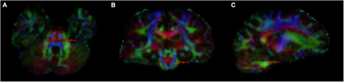
Materials and methods: DTI sequences was performed on PTN patients and healthy controls (HCs). Clinical data included affected side, disease course and visual analogue scale (VAS) were collected. Quantitative DTI variables such as FA, MD, AD and RD of the root entry/Exit zone (REZ) were measured and compared in PTN/HCs, affected/unaffected side, and pre/post operation groups. The PCoA was established to conduct overall differences between PTN group and the HCs.
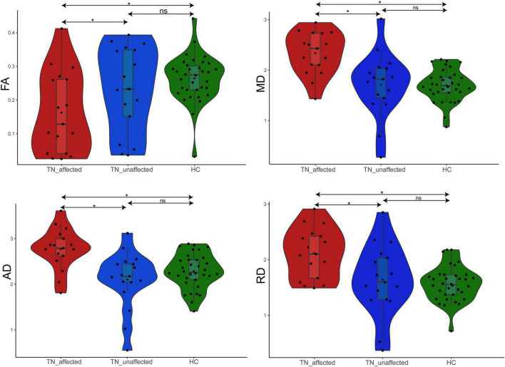
Results: A total of 17 patients with PTN (mean age 59.29 ± 8.53; 5 men) and 34 HCs (mean age 57.70 ± 6.37; 10 men) were included. Lower FA value of the affected side of PTN group was observed compared to the unaffected side and the HCs (p = 0.001), whereas the values of MD, AD and RD were significantly increased (p < 0.001). Moreover, the decrease of FA value was recovered post operation. PCoA results of the comprehensive indexes can significantly distinguish PTN group from HCs (r = 0.500, p < 0.001).
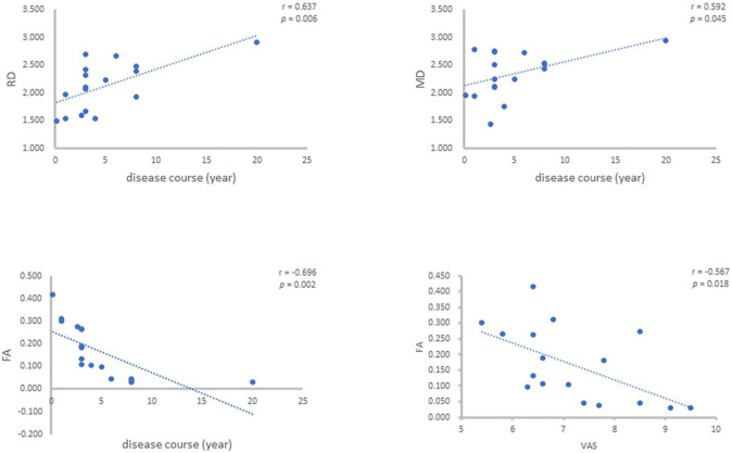
Conclusion: Quantitative variables derived from DTI in REZ had significantly different profiles between PTN patients and HCs, which were associated with VAS situation and the disease course of PTN. The comprehensive index established on DTI variables were of great potential to reveal the microstructure changes in PTN patients and predict the therapeutic effect.
Keywords: diffusion tensor imaging; magnetic resonance imaging; neurovascular compression (NVC); primary trigeminal neuralgia; root entry zone.
Copyright © 2023 Wang, Wang, Wu, Zhu, Wei, Li, Li, Chen and Chen.
Conflict of interest statement
The authors declare that the research was conducted in the absence of any commercial or financial relationships that could be construed as a potential conflict of interest.
Figures

Similar articles
-
Utility of DTI (Diffusion Tensor Imaging) Metrics to Study Microstructural Changes of Trigeminal Nerve in Patients with Trigeminal Neuralgia (TN).Neurol India. 2022 Jan-Feb;70(1):270-274. doi: 10.4103/0028-3886.338701. Neurol India. 2022. PMID: 35263894
-
Microstructural alterations in trigeminal neuralgia determined by diffusion tensor imaging are independent of symptom duration, severity, and type of neurovascular conflict.J Neurosurg. 2016 Mar;124(3):823-30. doi: 10.3171/2015.2.JNS142587. Epub 2015 Sep 25. J Neurosurg. 2016. PMID: 26406792
-
The timing of stereotactic radiosurgery for medically refractory trigeminal neuralgia: the evidence from diffusion tractography images.Acta Neurochir (Wien). 2018 May;160(5):977-986. doi: 10.1007/s00701-017-3449-9. Epub 2018 Feb 3. Acta Neurochir (Wien). 2018. PMID: 29397449
-
Magnetic resonance imaging contribution for diagnosing symptomatic neurovascular contact in classical trigeminal neuralgia: a blinded case-control study and meta-analysis.Pain. 2014 Aug;155(8):1464-1471. doi: 10.1016/j.pain.2014.04.020. Epub 2014 Apr 28. Pain. 2014. PMID: 24785270
-
Structural Magnetic Resonance Imaging Can Identify Trigeminal System Abnormalities in Classical Trigeminal Neuralgia.Front Neuroanat. 2016 Oct 19;10:95. doi: 10.3389/fnana.2016.00095. eCollection 2016. Front Neuroanat. 2016. PMID: 27807409 Free PMC article. Review.
Cited by
-
Assessment of Trigeminal Nerve Root Demyelination in Patients with Primary Trigeminal Neuralgia Using Macromolecular Proton Fraction Imaging.AJNR Am J Neuroradiol. 2025 Mar 4;46(3):602-610. doi: 10.3174/ajnr.A8545. AJNR Am J Neuroradiol. 2025. PMID: 40016130
-
The utility of diffusion tensor imaging in the assessment of trigeminal neuralgia pathophysiology and clinical outcome: a systematic review.Neurol Sci. 2025 Jun;46(6):2539-2554. doi: 10.1007/s10072-025-08019-8. Epub 2025 Feb 3. Neurol Sci. 2025. PMID: 39899218
-
Microstructural alterations of the trigeminal ganglion in chronic ocular surface pain patients: A diffusion MRI study.Neuroimage. 2025 Aug 15;317:121309. doi: 10.1016/j.neuroimage.2025.121309. Epub 2025 Jun 18. Neuroimage. 2025. PMID: 40554396 Free PMC article.
-
A study of brain function changes in patients with trigeminal neuralgia of different laterality based on rs-fMRI.J Oral Facial Pain Headache. 2025 Mar;39(1):148-156. doi: 10.22514/jofph.2025.015. Epub 2025 Mar 12. J Oral Facial Pain Headache. 2025. PMID: 40129433 Free PMC article.
References
LinkOut - more resources
Full Text Sources
Miscellaneous