. 2023 Mar 24:14:1070319.
doi: 10.3389/fpls.2023.1070319. eCollection 2023.

Phenotypic and histological analyses on the resistance of melon to Phelipanche aegyptiaca

Affiliations

Phenotypic and histological analyses on the resistance of melon to Phelipanche aegyptiaca

Xiaolei Cao et al. Front Plant Sci. .

Abstract

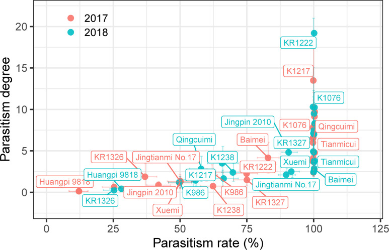
Melon (Cucumis melo L.) is an economically important crop in Xinjiang, China, but its production is constrained by the parasitic plant Phelipanche aegyptiaca that attaches to the roots of many crops and causes severe stunting and loss of yield. Rhizotron, pot, and field experiments were employed to evaluate the resistance of 27 melon cultivars to P. aegyptiaca. Then, the resistant and susceptible cultivars were inoculated with P. aegyptiaca from six populations to assess their resistance stability and broad spectrum. Further microscopic and histological analyses were used to clarify the resistance phenotypes and histological structure. The results showed that Huangpi 9818 and KR1326 were more resistant to P. aegyptiaca compared to other cultivars in the rhizotron, pot, and field experiments. In addition, compared to the susceptible cultivar K1076, Huangpi 9818 and KR1326 showed broad-spectrum resistance to six P. aegyptiaca populations. These two resistant cultivars had lower P. aegyptiaca biomass and fewer and smaller P. aegyptiaca attachments on their roots compared to susceptible cultivar K1076. KR1326 (resistant) and K1076 (susceptible) were selected to further study resistance phenotypes and mechanisms. Germination-inducing activity of root exudates and microscopic analysis showed that the resistance in KR1326 was not related to low induction of P. aegyptiaca germination. The tubercles of parasite on KR1326 were observed slightly brown at 14 days after inoculation (DAI), the necrosis and arrest of parasite development occurred at 23 DAI. Histological analysis of necrosis tubercles showed that the endophyte of parasite had reached host central cylinder, connected with host xylem, and accumulation of secretions and callose were detected in neighbouring cells. We concluded that KR1326 is an important melon cultivar for P. aegyptiaca resistance that could be used to expand the genetic basis of cultivated muskmelon for resistance to the parasite.

Keywords: Phelipanche aegyptiaca; histology; melon; necrosis; parasitic plants; resistance.


Conflict of interest statement

The authors declare that the research was conducted in the absence of any commercial or financial relationships that could be construed as a potential conflict of interest.

Figures

Figure 1
Figure 1
Percentages of P. aegyptiaca at different growth stages on roots of 27 melon cultivars at 35 DAI. S0, P. aegyptiaca seeds were not germinated; S1, seeds germinated but failed to attach to the melon roots; S3, seedlings had firmly adhered to or penetrated the host roots but had not formed a vascular connection; S4, tubercle stage, the parasite had formed a vascular connection to the host roots; S5, the “spider” stage (tubercles with secondary roots) ≤ 1cm. Vertical bars represent the mean ± SE, while letters represent mean separation at P ≤ 0.05 by Tukey’s HSD test in each growth stage.
Figure 2
Figure 2
Parasitism degree and parasitism rate of the different melon cultivars in pot experiments. Vertical and horizontal bars indicate standard errors of parasitism degree and rate, respectively (n = ~3–8, biological replicates). Only some representative melon cultivars are shown in the figure.
Figure 3
Figure 3
The resistance response of melon cultivars to P. aegyptiaca at the experimental station and Majiaping field sites. K1076, Xuemi, Baimei, K1217, Jingtianmi No.17, Huangpi 9818, and KR1326 were used in the field trials. (A) The number of emerged P. aegyptiaca plants per cultivar for the experimental station in 2018 and Majiaping in 2019 and 2021. Data presented are means ± SE. The letters above each bar indicate significantly different (P ≤ 0.05) groups after Tukey’s HSD test. (B) Contrasting P. aegyptiaca infection levels in the P. aegyptiaca screening trial at Majiaping (July 2021) replicate 3; sub-plots, representing cultivars are delimited by white lines. (C) The growth of Xuemi, KR1326, and K1076 at Majiaping (July 2021). Red arrows represent the emerged P. aegyptiaca shoots. (D, F) Single melon mass (Kg) per cultivar for the experimental station in 2018 and Majiaping in 2021. (E, G) Melon yield (t ha-1) per cultivar for the experimental station in 2018 and Majiaping in 2021. Data presented are means ± SE. NS, *, **, *** indicate significant differences at P > 0.05, P < 0.05, P < 0.01, and P < 0.001 respectively, by Student’s t test within a variety.
Figure 4
Figure 4
Development and biomass of six P. aegyptiaca populations attached to the roots of KR1326, Huangpi9818, and K1076 at 60 DAI. (A–D) The number of P. aegyptiaca at S4, S5, S6, S7 stages per host plant. (E) Total number of underground attachments per host plant (UAN). (F) Total number of P. aegyptiaca attachments per plant (TAN). (G) Fresh weight of total attachments per host plant. (H) Dry weight of total attachments per host plant. Vertical bars represent the mean ± SE, while letters represent mean separations at P ≤ 0.05 by Tukey’s HSD test (n = 10 biological replicates).
Figure 5
Figure 5
Effects of six P. aegyptiaca populations on the growth of different resistant melon cultivars. (A) Effects of P. aegyptiaca on melon total aboveground parts dry weight (GDW). (B) Effects of P. aegyptiaca on melon root dry weight (RDW). (C) Effects of P. aegyptiaca on melon plant height (PH). (D) Effects of P. aegyptiaca on melon stem diameter (SD). (E) Growth of melon after inoculation with or without broomrape from six populations. Vertical bars represent the mean ± SE, while letters represent mean separations at P ≤ 0.05 by Tukey’s HSD test (n = 10 biological replicates).
Figure 6
Figure 6
Differences in root exudate of susceptible (K1076) and resistant (KR1326) melon cultivars for germination induction of P. aegyptiaca (A) and O. cumana (B) seeds and differences in contents of 5-DS and Strigol in roots (C). The crude extract of K1076 and KR1326 root exudates was dissolved with 1 ml isopropanol and then diluted with sterile water 4, 10, 1 × 10², 1 × 10³, 1 × 10⁴, 1 × 10⁵, and 1 × 10⁶ times (n ≥ 3 replicates). Seeds were treated with isopropanol diluted with sterile water at 4, 10, 1 × 10², 1 × 10³, 1 × 10⁴, 1 × 10⁵, and 1 × 10⁶ times as negative controls. Seeds treated with or without GR24 at 1 × 10⁻⁷ M were always included as positive and negative controls, respectively. (C) Amounts of 5-DS and Strigol (ng/g) in roots (2g) of K1076 and KR1326 obtained in hydroponics (n=3 biological replicates). (D) 1 × 10²-time concentrated root exudates from K1076 and KR1326 and GR24 induced germination of P. aegyptiaca and O. cumana. Experiments were repeated at least three times. Scale bar, 500 μm. Vertical bars represent the mean ± SE and P value was conducted by Student t test.
Figure 7
Figure 7
Differential response of K1076 and KR1326 to parasitism by P. aegyptiaca. (A) Representative photographs illustrating the phenotypic response of K1076, a susceptible cultivar, and KR1326 a resistant cultivar, to parasitism by P. aegyptiaca. Shown are the appearance of broomrape and the attachment sites at 7, 9, 16, and 23 DAI (the broomrape was in S2, S3, S4, and S5 stages, respectively). Bars = 200 μm. (B) Measured percentage of each different phenotypic event category during the interaction of P. aegyptiaca with resistant (KR1326) and susceptible (K1076) melon roots at 11, 16, and 23 DAI (n = 5~10). The abbreviation of the phenotypic event categories are as follows: S3, seeds had firmly adhered to or penetrated the host roots but had not formed a vascular connection; S4H, healthy tubercles; S4B, tubercle slight discoloration (black arrow); S4DB, tubercle severely browning or necrosis (red arrow); S5H, broomrape is healthy in the “spider” stage; S5DB, broomrape shows severe browning or necrosis in the “spider” stage. Data presented are means ± SE. *, **, *** indicate significant differences at P < 0.05, P < 0.01, and P < 0.001 respectively, by Student’s t test within a variety.
Figure 8
Figure 8
Transverse sections through the root and P. aegyptiaca attachment in compatible and incompatible interactions at 16 and 23 DAI. (A, E) Light micrograph of a cross-section in a healthy broomrape on K1076 (16 and 23 DAI). (I) Detail of (E). (B, F, J) The same as (A, E, I) under fluorescence microscopy (450–490 nm). (C, G) Light micrograph of a cross-section in a slightly discolored and severely browned tubercle on KR1326 (16 and 23 DAI). (K) Detail of (G). (D, H, L) The same as (C, G, K) under fluorescence microscopy (450–490 nm). p, parasite (P. aegyptiaca); px, parasite xylem; hx, host xylem; he, host endodermis; hc, host root cortex; en, endophyte. The red arrow indicates the young stem meristem of the broomrape; the white arrow indicates the secondary root meristem; the white triangle indicates autofluorescence of the host xylem; the red triangle indicates autofluorescence of the xylem of the broomrape; the black triangle indicates the accumulation of secondary metabolites in the host cell wall; the red box indicates parasite xylem, and a white border indicates parenchyma cells of the parasite. Bars = 200 μm.
Figure 9
Figure 9
Cross-sections of incompatible and compatible interactions of P. aegyptiaca on KR1326 and K1076 stained with aniline blue at 16 DAI. Callose deposition shows as a blue-white fluorescence (arrows). (A, B, E, F) Light micrographs of fresh hand-cut sections of noninfected (A, B) and infected (E, F) with broomrape roots of KR1326. (C, D, G, H) The same as (A, B, E, F) observed under fluorescence (340–380 nm). (H) Magnification of the square box in (G). (I, J, M, N) Light micrograph of a fresh hand-cut section of a noninfected (I, J) and infected (M, N) with broomrape roots of K1076. (K, L, O, P) The same as (I, J, M, N) observed under fluorescence (340–380 nm). (P) Magnification of the square box in O. Bars = 100 μm. h, host; p, parasite (P. aegyptiaca); hx, host xylem; en, endophyte.

References

    1. Aly R. (2013). Trafficking of molecules between parasitic plants and their hosts. Weed. Res. 53, 231–241. doi: 10.1111/wre.12025 - DOI
    1. Anderson I. C., Cairney J. W. G. (2004). Diversity and ecology of soil fungal communities: Increased understanding through the application of molecular techniques. Environ. Microbio. 6, 769–779. doi: 10.1111/j.1462-2920.2004.00675.x - DOI - PubMed
    1. Bai J., Wei Q., Shu J., Gan Z., Li B., Yan D., et al. . (2020). Exploration of resistance to Phelipanche aegyptiaca in tomato. Pest Manage. Sci. 76, 3806–3821. doi: 10.1002/ps.5932 - DOI - PubMed
    1. Bao Y. Z., Yao Z. Q., Cao X. L., Peng J. F., Xu Y., Chen M. X., et al. . (2017). Transcriptome analysis of Phelipanche aegyptiaca seed germination mechanisms stimulated by fluridone, TIS108, and GR24. PloS One 12, e0187539. doi: 10.1371/journal.pone.0187539 - DOI - PMC - PubMed
    1. Bin L., Wang C., Liu Z., He W., Zhao D., Fang Y. Y., et al. . (2022). Geographical origin traceability of muskmelon from xinjiang province using stable isotopes and multi-elements with chemometrics. J. Food Compos. Anal. 106, 104320. doi: 10.1016/j.jfca.2021.104320 - DOI

LinkOut - more resources