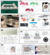
Recent development of microfluidics-based platforms for respiratory virus detection
- PMID: 37035101
- PMCID: PMC10076069
- DOI: 10.1063/5.0135778
Recent development of microfluidics-based platforms for respiratory virus detection
Abstract
With the global outbreak of SARS-CoV-2, the inadequacies of current detection technology for respiratory viruses have been recognized. Rapid, portable, accurate, and sensitive assays are needed to expedite diagnosis and early intervention. Conventional methods for detection of respiratory viruses include cell culture-based assays, serological tests, nucleic acid detection (e.g., RT-PCR), and direct immunoassays. However, these traditional methods are often time-consuming, labor-intensive, and require laboratory facilities, which cannot meet the testing needs, especially during pandemics of respiratory diseases, such as COVID-19. Microfluidics-based techniques can overcome these demerits and provide simple, rapid, accurate, and cost-effective analysis of intact virus, viral antigen/antibody, and viral nucleic acids. This review aims to summarize the recent development of microfluidics-based techniques for detection of respiratory viruses. Recent advances in different types of microfluidic devices for respiratory virus diagnostics are highlighted, including paper-based microfluidics, continuous-flow microfluidics, and droplet-based microfluidics. Finally, the future development of microfluidic technologies for respiratory virus diagnostics is discussed.
© 2023 Author(s).
Conflict of interest statement
The authors have no conflicts to disclose.
Figures


Similar articles
-
Microfluidic-based virus detection methods for respiratory diseases.Emergent Mater. 2021;4(1):143-168. doi: 10.1007/s42247-021-00169-7. Epub 2021 Mar 25. Emergent Mater. 2021. PMID: 33786415 Free PMC article. Review.
-
Conventional and microfluidic methods for airborne virus isolation and detection.Colloids Surf B Biointerfaces. 2021 Oct;206:111962. doi: 10.1016/j.colsurfb.2021.111962. Epub 2021 Jul 2. Colloids Surf B Biointerfaces. 2021. PMID: 34352699 Free PMC article. Review.
-
Microfluidics-based strategies for molecular diagnostics of infectious diseases.Mil Med Res. 2022 Mar 18;9(1):11. doi: 10.1186/s40779-022-00374-3. Mil Med Res. 2022. PMID: 35300739 Free PMC article. Review.
-
Recent Advances in Molecular and Immunological Diagnostic Platform for Virus Detection: A Review.Biosensors (Basel). 2023 Apr 19;13(4):490. doi: 10.3390/bios13040490. Biosensors (Basel). 2023. PMID: 37185566 Free PMC article. Review.
-
Development of automated microfluidic immunoassays for the detection of SARS-CoV-2 antibodies and antigen.J Immunol Methods. 2024 Jan;524:113586. doi: 10.1016/j.jim.2023.113586. Epub 2023 Nov 30. J Immunol Methods. 2024. PMID: 38040191
Cited by
-
Open-source and low-cost miniature microscope for on-site fluorescence detection.HardwareX. 2024 Jun 15;19:e00545. doi: 10.1016/j.ohx.2024.e00545. eCollection 2024 Sep. HardwareX. 2024. PMID: 39006472 Free PMC article.
-
Impact of Nested Multiplex Polymerase Chain Reaction Assay in the management of pediatric patients with acute respiratory tract infections: a single center experience.Infez Med. 2023 Dec 1;31(4):539-552. doi: 10.53854/liim-3104-13. eCollection 2023. Infez Med. 2023. PMID: 38075410 Free PMC article.
-
Lateral Flow Biosensor for On-Site Multiplex Detection of Viruses Based on One-Step Reverse Transcription and Strand Displacement Amplification.Biosensors (Basel). 2024 Feb 17;14(2):103. doi: 10.3390/bios14020103. Biosensors (Basel). 2024. PMID: 38392022 Free PMC article.
-
Ultrasensitive detection of intact SARS-CoV-2 particles in complex biofluids using microfluidic affinity capture.Sci Adv. 2025 Jan 10;11(2):eadh1167. doi: 10.1126/sciadv.adh1167. Epub 2025 Jan 10. Sci Adv. 2025. PMID: 39792670 Free PMC article.
References
-
- Boncristiani H. F., Criado M. F., and Arruda E., Encyclopedia Microbiol. 4, 500–518 (2009). 10.1016/B978-012373944-5.00314-X - DOI
-
- Patterson K. D. and Pyle G. F., Bull. Hist. Med. 65, 4–21 (1991); available at http://www.jstor.org/stable/44447656. - PubMed
LinkOut - more resources
Full Text Sources
Miscellaneous
