. 2023 Mar 23:13:1145379.
doi: 10.3389/fonc.2023.1145379. eCollection 2023.

YB1 participated in regulating mitochondrial activity through RNA replacement

Affiliations

YB1 participated in regulating mitochondrial activity through RNA replacement

Weipeng Gong et al. Front Oncol. .

Abstract

As a relic of ancient bacterial endosymbionts, mitochondria play a central role in cell metabolism, apoptosis, autophagy, and other processes. However, the function of mitochondria-derived nucleic acids in cellular signal transduction has not been fully elucidated. Here, our work has found that Y-box binding protein 1 (YB1) maintained cellular autophagy at a moderate level to inhibit mitochondrial oxidative phosphorylation. In addition, mitochondrial RNA was leaked into cytosol under starvation, accompanied by YB1 mitochondrial relocation, resulting in YB1-bound RNA replacement. The mRNAs encoded by oxidative phosphorylation (OXPHOS)-associated genes and oncogene HMGA1 (high-mobility group AT-hook 1) were competitively replaced by mitochondria-derived tRNAs. The increase of free OXPHOS mRNAs released from the YB1 complex enhanced mitochondrial activity through facilitating translation, but the stability of HMGA1 mRNA was impaired without the protection of YB1, both contributing to breast cancer cell apoptosis and reactive oxygen species production. Our finding not only provided a new potential target for breast cancer therapy but also shed new light on understanding the global landscape of cellular interactions between RNA-binding proteins and different RNA species.

Keywords: HMGA1; YB1; apoptosis; autophagy; mitochondria.


Conflict of interest statement

The authors declare that the research was conducted in the absence of any commercial or financial relationships that could be construed as a potential conflict of interest.

Figures

Figure 1
Figure 1
The upregulation of YB1 in breast cancerous tissues was associated with poor clinical outcomes. (A) Relative expression of YB1 in normal or cancerous tissues of patients with breast cancer. The expression level of YB1 in metastatic cancerous tissues was specifically elevated compared with normal or non-metastatic tissues (https://tnmplot.com). (B) Expression level of YB1 in breast cancer patients under different clinical stages (http://gent2.appex.kr/gent2/). (C) The relationship between YB1 expression and breast cancer types (http://gent2.appex.kr/gent2/). (D-F) Kaplan–Meier survival rates for patients with breast cancer stratified by YB1 expression. The survival rates for patients were plotted separately according to YB1 expression at the mRNA (D) or protein level (E) (http://kmplot.com/analysis/). The most remarkable difference emerged in patients with basal-like/triple-negative breast cancer (F). (G) Immunohistochemical analysis of YB1 in solid tumors. Paired paracarcinoma and carcinoma tissues were excised from the same patient with breast cancer.
Figure 2
Figure 2
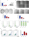
YB1 depletion inhibited breast cancer cell proliferation and migration. (A, B) Knockdown efficiency of YB1-targeted shRNAs in breast cancer cells (MDA-MB-231) at the mRNA level and protein level. β-Tubulin was used as a loading control. The experiment was repeated three times, **P < 0.01. (C) Colony formation assay was performed to investigate the influence of YB1 depletion on cancer cell proliferation. The experiment was repeated three times, **P < 0.01. (D, E) The function of YB1 on cell migration ability was investigated using wound-healing and Transwell assays. Scale bar, 100 μm. (F) YB1 expression silence induced obvious S-stage cell-cycle arrest in breast cancer cells. The cell cycle was investigated through flow cytometry. (G) The influence of YB1 knockdown on cell apoptosis detected by flow cytometry. Experiments were repeated three times.
Figure 3
Figure 3
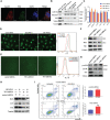
YB1 participated in balancing autophagy and cell apoptosis. (A) The co-localization of YB1 (green) with mitochondria (red) in breast cancer cells. Mitochondria were marked with TOM20 (red), and nuclei were stained with DAPI. Scale bars, 5 μm. (B) The protein level of YB1 in different mitochondrial fractions. Digitonin dose-dependently dissolved the mitochondrial outer membrane. The residual proteins were digested with protease K NDUFA9, inner membrane protein marker; LRPPRC, mitochondrial matrix localized protein. (C) Relative expressions of mitochondrial genome-encoded genes. The experiment was repeated three times. **p < 0.01. (D) The activity of mitochondria was investigated using flow cytometry. Mitochondria were labeled with MitoTracker dye; the fluorescence signal intensity was analyzed. Scale bar, 10 μm. (E) The influence of YB1 knockdown on cellular reactive oxygen species. Scale bar, 10 μm. (F) YB1 knockdown reduced the cellular basic autophagy level in breast cancer cells. (G) The mTOR signaling pathway was activated in cells with deleted YB1. (H, I) YB1-silenced expression failed to induce autophagy under starvation but significantly increased cell apoptosis.
Figure 4
Figure 4
YB1 interacted with mitochondrial tRNA. (A, B) RNA-associated immunoprecipitation (RIP) assays. RIP was conducted in MDA-MB-231 cells to sediment the YB1–RNA complex; rabbit IgG was used as a negative control. The contents of immunoprecipitate were analyzed by SDS-PAGE with Coomassie staining (A) or Western blot (B). (C) Detection of mitochondrial RNA in the YB1-immunoprecipitated complex. The RNAs co-immunoprecipitated with YB1 proteins were isolated and detected using quantitative real-time PCR. YB1 distribution was detected using the mitochondrial fraction (D) and immunofluorescence (E); scale bar, 10 μm. (F) Serum-free starvation treatment increased the direct interaction between mitochondrial RNA and YB1 protein. n = 3, **p < 0.01, *p < 0.05. (G) MT-TF distribution detection using FISH. After starvation treatment, more MT-TF (red) were localized in the outside of mitochondria. Scale bar, 10 μm. (H) A YB1–RNA interaction model was built using the online SWISS-MODEL.
Figure 5
Figure 5
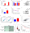
YB1 maintained the stability of HMGA1 mRNA. (A) Detection of oncogenes in the YB1-bound RNA library through RIP assays. (B, C) YB1 knockdown significantly downregulated HMGA1 expression at both mRNA level and protein level. (D) Serum-free starvation impaired YB1 binding ability with HMGA1 mRNA. n = 3,**p < 0.01. (E) Influence of YB1 knockdown on HMGA1 mRNA degradation. (F) The expression of HMGA1 was silenced using siRNA. (G) Influence of HMGA1 knockdown on cell apoptosis. n = 3, **p < 0.01. (H) Correlation analysis of YB1 expression with HMGA1 expression in breast cancer tissues. Data were downloaded from website http://timer.comp-genomics.org. (I) Immunohistochemical analysis of HMGA1 in paired breast carcinoma and paracarcinoma tissues. (J) Analysis of HMGA1 expression in breast cancer tissues and normal tissues through referring to an online database (http://gepia.cancer-pku.cn/). *p < 0.05. (K) High expression of HMGA1 in breast cancer tissues predicts adverse clinical survival outcomes (http://kmplot.com/analysis/).
Figure 6
Figure 6
YB1 inhibited OXPHOS gene translation. (A) The influence of YB1 knockdown on mitochondrial oxidative phosphorylation gene (NDUFA9 and SDHB) expressions at the mRNA level. n = 3, **p < 0.01. (B) YB1 expression silence enhanced NDUFA9 and SDHB protein expressions through posttranscriptional regulation. (C) The influence of starvation on DNUFA9 and SDHB expressions was investigated through Western blot. (D) YB1 directly interacted with OXPHOS mRNA. The interaction between YB1 protein and OXPHOS mRNA (NDUFA9 and SDHB) was demonstrated using RIP assay. n = 3, **p < 0.01. (E) Serum-free starvation impaired YB1 binding ability with OXPHOS mRNAs. n = 3, *p < 0.05. (F) The protein levels of NDUFA9 and SDHB were investigated in breast cancer cells with or without YB1 depletion. MG132 was used to block the proteasome-mediated protein degradation pathway. (G) The protein degradation rates of NDUFA9 and SDHB were investigated. CHX was used to block the elongation phase of eukaryotic translation. (H) The influence of MT-TF overexpression (MT-TF OE) on breast cancer proliferation was detected using MTT assay. (I) The migration ability of breast cancer was inhibited after MT-TF overexpression. Scale bar, 100 μm. (J) The expressions of NDUFA9, SDHB, and HMGA1 were investigated in breast cancer cells transfected with MT-TF.
Figure 7
Figure 7
YB1 promoted tumorigenesis of breast cancer cells. (A) The statistics of xenograft volume in nude mice at different times. Breast cancer cells with different YB1 expression levels were subcutaneously injected into nude mice, and the volumes of tumors were monitored at different times. n = 5, *p < 0.05, **p < 0.01. (B) The representative pictures of solid tumors collected from nude mice. (C) Solid tumor weight analysis. n = 5, **p < 0.01. (D) Immunohistochemical analysis of YB-1-regulated proteins in breast cancer xenografts. Specific antibodies were used to detect each protein (brown), and nuclei were stained with hematoxylin (blue). (E) The levels of YB1-regulated proteins in xenografts were investigated through Western blot. (F) Proposed working model for the role of starvation-induced YB1-bound RNA replacement by mitochondrial RNA in regulating cell autophagy and apoptosis in breast cancer cells.

Similar articles

Cited by

References

    1. Silva J, Aivio S, Knobel PA, Bailey LJ, Casali A, Vinaixa M, et al. . EXD2 governs germ stem cell homeostasis and lifespan by promoting mitoribosome integrity and translation. Nat Cell Biol (2018) 20(2):162–74. doi: 10.1038/s41556-017-0016-9 - DOI - PubMed
    1. Kauppila T, Kauppila J, Larsson NG. Mammalian mitochondria and aging: An update. Cell Metab (2017) 25(1):57–71. doi: 10.1016/j.cmet.2016.09.017 - DOI - PubMed
    1. Zhao Q, Liu J, Deng H, Ma R, Liao JY, Liang H, et al. . Targeting mitochondria-located circRNA SCAR alleviates NASH via reducing mROS output. Cell (2020) 183(1):76–93.e22. doi: 10.1016/j.cell.2020.08.009 - DOI - PubMed
    1. Rambold AS, Pearce EL. Mitochondrial dynamics at the interface of immune cell metabolism and function. Trends Immunol (2018) 39(1):6–18. doi: 10.1016/j.it.2017.08.006 - DOI - PubMed
    1. Garcia-Heredia JM, Carnero A. Role of mitochondria in cancer stem cell resistance. Cells (2020) 9(7):1693. doi: 10.3390/cells9071693 - DOI - PMC - PubMed