

The forgotten key players in rheumatoid arthritis: IL-8 and IL-17 - Unmet needs and therapeutic perspectives
- PMID: 37035302
- PMCID: PMC10073515
- DOI: 10.3389/fmed.2023.956127
The forgotten key players in rheumatoid arthritis: IL-8 and IL-17 - Unmet needs and therapeutic perspectives
Abstract
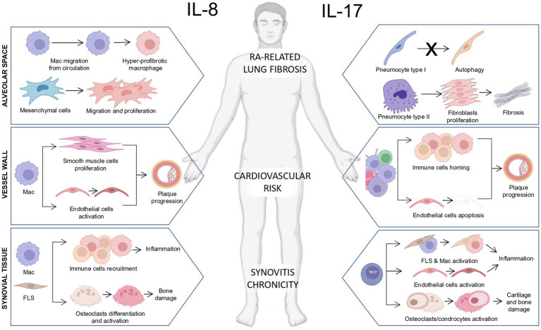
Despite the relevant advances in our understanding of the pathogenetic mechanisms regulating inflammation in rheumatoid arthritis (RA) and the development of effective therapeutics, to date, there is still a proportion of patients with RA who do not respond to treatment and end up progressing toward the development of joint damage, extra-articular complications, and disability. This is mainly due to the inter-individual heterogeneity of the molecular and cellular taxonomy of the synovial membrane, which represents the target tissue of RA inflammation. Tumor necrosis factor alpha (TNFα) and interleukin-6 (IL-6) are crucial key players in RA pathogenesis fueling the inflammatory cascade, as supported by experimental evidence derived from in vivo animal models and the effectiveness of biologic-Disease Modifying Anti-Rheumatic Drugs (b-DMARDs) in patients with RA. However, additional inflammatory soluble mediators such as IL-8 and IL-17 exert their pathogenetic actions promoting the detrimental activation of immune and stromal cells in RA synovial membrane, tendons, and extra-articular sites, as well as blood vessels and lungs, causing extra-articular complications, which might be excluded by the action of anti-TNFα and anti-IL6R targeted therapies. In this narrative review, we will discuss the role of IL-8 and IL-17 in promoting inflammation in multiple biological compartments (i.e., synovial membrane, blood vessels, and lung, respectively) in animal models of arthritis and patients with RA and how their selective targeting could improve the management of treatment resistance in patients.
Keywords: chronic pain; interleukin-17; interleukin-8; organ damage; rheumatoid arthritis.
Copyright © 2023 Gremese, Tolusso, Bruno, Perniola, Ferraccioli and Alivernini.
Conflict of interest statement
The authors declare that the research was conducted in the absence of any commercial or financial relationships that could be construed as a potential conflict of interest.
Figures
Similar articles
-
What have we learnt from the inhibition of IL-6 in RA and what are the clinical opportunities for patient outcomes?Ther Adv Musculoskelet Dis. 2024 Oct 21;16:1759720X241283340. doi: 10.1177/1759720X241283340. eCollection 2024. Ther Adv Musculoskelet Dis. 2024. PMID: 39444594 Free PMC article. Review.
-
Periodontal Disease as a Risk Factor for Rheumatoid Arthritis: A Systematic Review.JBI Libr Syst Rev. 2012;10(42 Suppl):1-12. doi: 10.11124/jbisrir-2012-288. JBI Libr Syst Rev. 2012. PMID: 27820156
-
Mast cell activation and its relation to proinflammatory cytokine production in the rheumatoid lesion.Arthritis Res. 2000;2(1):65-74. doi: 10.1186/ar70. Arthritis Res. 2000. PMID: 11219391 Free PMC article.
-
[Rheumatoid arthritis: new developments in the pathogenesis with special reference to synovial fibroblasts].Z Rheumatol. 2001 Oct;60(5):309-18. doi: 10.1007/s003930170030. Z Rheumatol. 2001. PMID: 11759230 Review. German.
-
Differential expression of pro-inflammatory cytokines IL-15Ralpha, IL-15, IL-6 and TNFalpha in synovial fluid from rheumatoid arthritis patients.BMC Musculoskelet Disord. 2015 Mar 12;16:51. doi: 10.1186/s12891-015-0516-3. BMC Musculoskelet Disord. 2015. PMID: 25879761 Free PMC article.
Cited by
-
ColMA-based bioprinted 3D scaffold allowed to study tenogenic events in human tendon stem cells.Bioeng Transl Med. 2024 Oct 30;10(1):e10723. doi: 10.1002/btm2.10723. eCollection 2025 Jan. Bioeng Transl Med. 2024. PMID: 39801753 Free PMC article.
-
Synoviocytes assist in modulating the effect of Ross River virus infection in micromass-cultured primary human chondrocytes.J Med Microbiol. 2024 Jul;73(7):001859. doi: 10.1099/jmm.0.001859. J Med Microbiol. 2024. PMID: 39028255 Free PMC article.
-
How to Distinguish Non-Inflammatory from Inflammatory Pain in RA?Curr Rheumatol Rep. 2024 Dec;26(12):403-413. doi: 10.1007/s11926-024-01159-4. Epub 2024 Aug 9. Curr Rheumatol Rep. 2024. PMID: 39120749 Free PMC article. Review.
-
Exploring perinatal mesenchymal stromal cells as a potential therapeutic strategy for rheumatoid arthritis.Heliyon. 2024 Dec 21;11(1):e41438. doi: 10.1016/j.heliyon.2024.e41438. eCollection 2025 Jan 15. Heliyon. 2024. PMID: 39811302 Free PMC article. Review.
-
Aqueous Humor Cytokine Profiling Reveals Distinct Roles for Serum Amyloid A, Interleukin-8, and Endothelin-1 in Pseudoexfoliation Syndrome and Glaucoma.Int J Mol Sci. 2025 Feb 10;26(4):1461. doi: 10.3390/ijms26041461. Int J Mol Sci. 2025. PMID: 40003925 Free PMC article.
References
-
- Bresnihan B, Flanagan AM, Firenstein GS. Synovium In: Firestein G, editor. Kelley's Textbook of Rheumatology. Vol. 1. 9th edn, Elsevier. (2013). 20–32.
Publication types
LinkOut - more resources
Full Text Sources
Research Materials