

Analysis on the genome of a teschovirus type 1 isolates with swine diarrhea
- PMID: 37035382
- PMCID: PMC10073753
- DOI: 10.1016/j.heliyon.2023.e14710
Analysis on the genome of a teschovirus type 1 isolates with swine diarrhea
Abstract
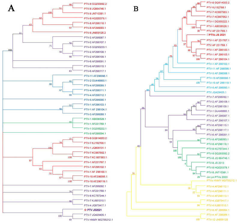
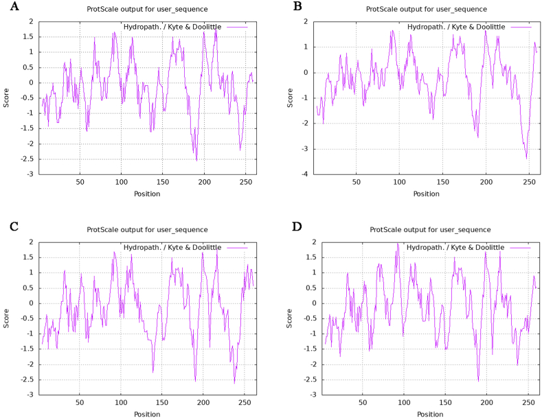
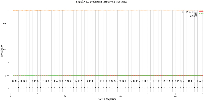
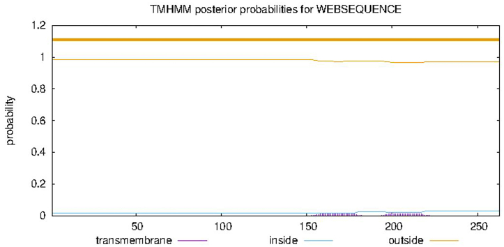
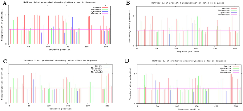
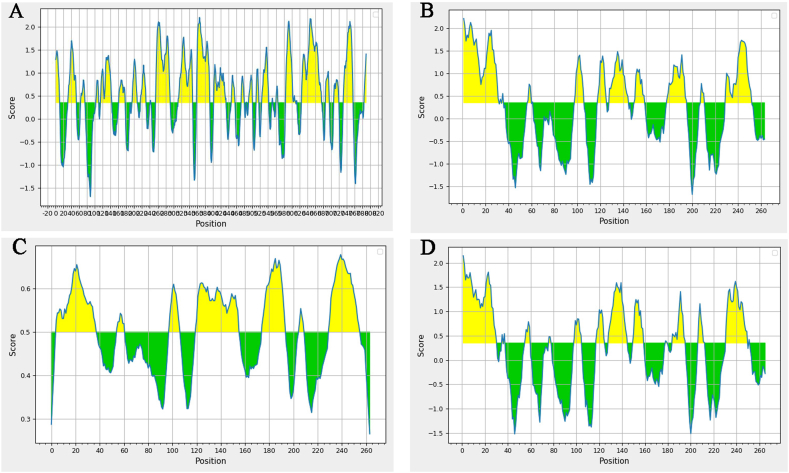
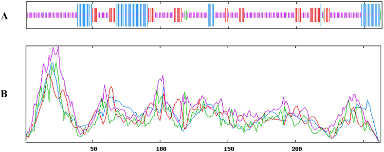
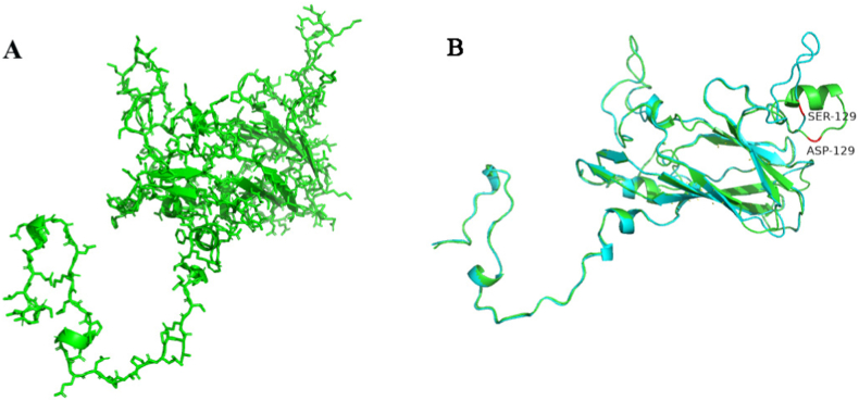
Porcine Teschoviruses (PTVs) are associated with polioencephalomyelitis and various diseases, including reproductive and gastrointestinal disorders of pigs and wild boars, but rarely detected in the feces of pigs. In this study, a sample of swine diarrhea that tested positive for PTVs is subjected to high-throughput sequencing. The viral genome was 7221 nucleotides (nt) in length, which was consisted of twelve genes. Phylogenetic analysis showed and it was closely related to the PTV-HNMY(MG755212.1). The nucleotide homology of VP1 gene of PTVs JS2021 with PTV-1AF 296102.1 reached 82.97%, belonging to a branch of PTV-1 serotype. The nucleotide homology of VP1 protein with other serotypes of PTV is quite different from that of other serotypes of PTV. Bioinformatics analysis showed that PTVs have four capsid proteins, namely VP1, VP2, VP3 and VP4. The VP1 encodes a 29 kDa protein, which is the main protective antigen, a theoretical isoelectric point of 6.73, no transmembrane domain, no signal peptide and potential phosphorylation site. The VP1 protein is an unstable hydrophilic intracellular protein, which contains four secondary structures: irregular curl (c), extended chain (e), α-helix (h) and β-folded (t). The tertiary structure is heart-shaped and has multiple B cell epitopes. By analyzing the tertiary structure, we found that the amino acid at position 129 of VP1 mutated and reduction a larger alpha helix. This may lead to the main cause of piglet diarrhea. These findings enriched our knowledge of the viruses in the role of swine diarrhea, and help to develop an effective strategy for disease prevention and control.
Keywords: Diarrhea; High-throughput sequencing; PTVs; Phylogenetic analysis; VP1.
©2023PublishedbyElsevierLtd.
Conflict of interest statement
The authors declare that they have no known competing financial interests or personal relationships that could have appeared to influence the work reported in this paper.
Figures
Similar articles
-
Metagenomic identification and sequence analysis of a Teschovirus A-related virus in porcine feces in Japan, 2014-2016.Infect Genet Evol. 2018 Dec;66:210-216. doi: 10.1016/j.meegid.2018.10.004. Epub 2018 Oct 11. Infect Genet Evol. 2018. PMID: 30316885
-
Epidemiology and molecular characterization of Porcine teschovirus in Hunan, China.Transbound Emerg Dis. 2018 Apr;65(2):480-490. doi: 10.1111/tbed.12728. Epub 2017 Oct 16. Transbound Emerg Dis. 2018. PMID: 29034572
-
Identification of a novel porcine Teschovirus 2 strain as causative agent of encephalomyelitis in suckling piglets with high mortality in China.BMC Vet Res. 2023 Jan 4;19(1):2. doi: 10.1186/s12917-022-03549-1. BMC Vet Res. 2023. PMID: 36597091 Free PMC article.
-
Isolation and genetic characteristics of a neurotropic teschovirus variant belonging to genotype 1 in northeast China.Arch Virol. 2021 May;166(5):1355-1370. doi: 10.1007/s00705-021-04994-3. Epub 2021 Mar 12. Arch Virol. 2021. PMID: 33709216
-
Validation of rt-PCR assays for molecular characterization of porcine teschoviruses and enteroviruses.J Vet Med B Infect Dis Vet Public Health. 2006 Aug;53(6):257-65. doi: 10.1111/j.1439-0450.2006.00955.x. J Vet Med B Infect Dis Vet Public Health. 2006. PMID: 16907956
Cited by
-
Development and implementation of a TaqMan triplex real-time PCR assay for concurrent detection of pseudorabies virus, porcine teschovirus 1, and Streptococcus suis 2.Front Vet Sci. 2025 Jun 18;12:1589175. doi: 10.3389/fvets.2025.1589175. eCollection 2025. Front Vet Sci. 2025. PMID: 40607354 Free PMC article.
-
A Quadruplex RT-qPCR for the Detection of Porcine Sapelovirus, Porcine Kobuvirus, Porcine Teschovirus, and Porcine Enterovirus G.Animals (Basel). 2025 Mar 31;15(7):1008. doi: 10.3390/ani15071008. Animals (Basel). 2025. PMID: 40218401 Free PMC article.
References
-
- Yamada M., Kaku Y., Nakamura K., Yoshii M., Yamamoto Y., Miyazaki A., Tsunemitsu H., Narita M. Immunohistochemical detection of porcine teschovirus antigen in the formalin-fixed paraffin-embedded specimens from pigs experimentally infected with porcine teschovirus. J. Vet. Med. 2007;54(10):571–574. - PubMed
LinkOut - more resources
Full Text Sources
Research Materials
