. 2022 Apr;24(4):82.
doi: 10.1007/s11051-022-05463-x. Epub 2022 Apr 13.

Photosensitized co-generation of nitric oxide and singlet oxygen Enhanced toxicity against ovarian cancer cells

Affiliations

Photosensitized co-generation of nitric oxide and singlet oxygen Enhanced toxicity against ovarian cancer cells

Pedro Sanchez-Cruz et al. J Nanopart Res. 2022 Apr.

Abstract

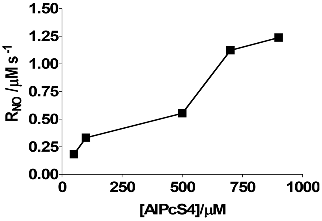
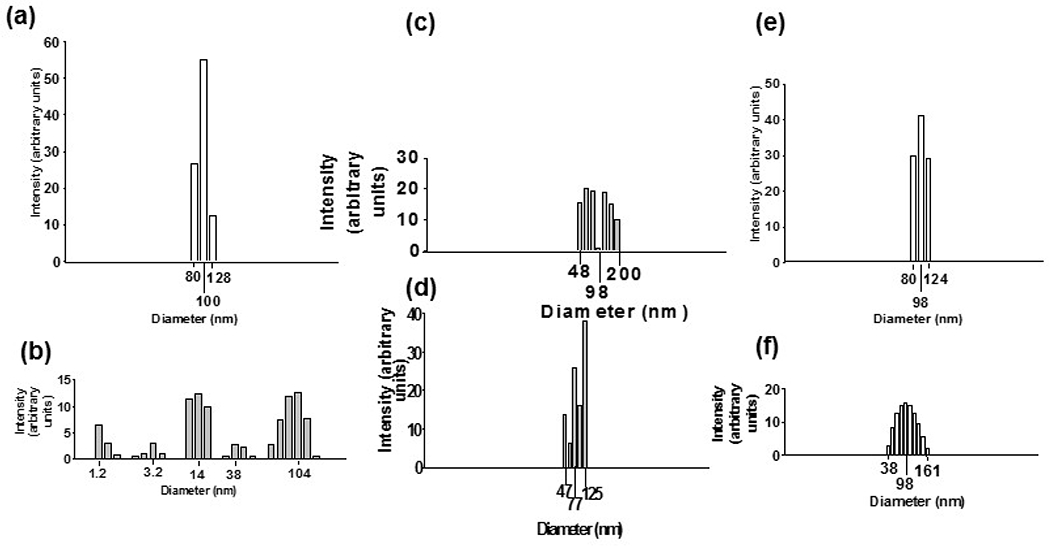
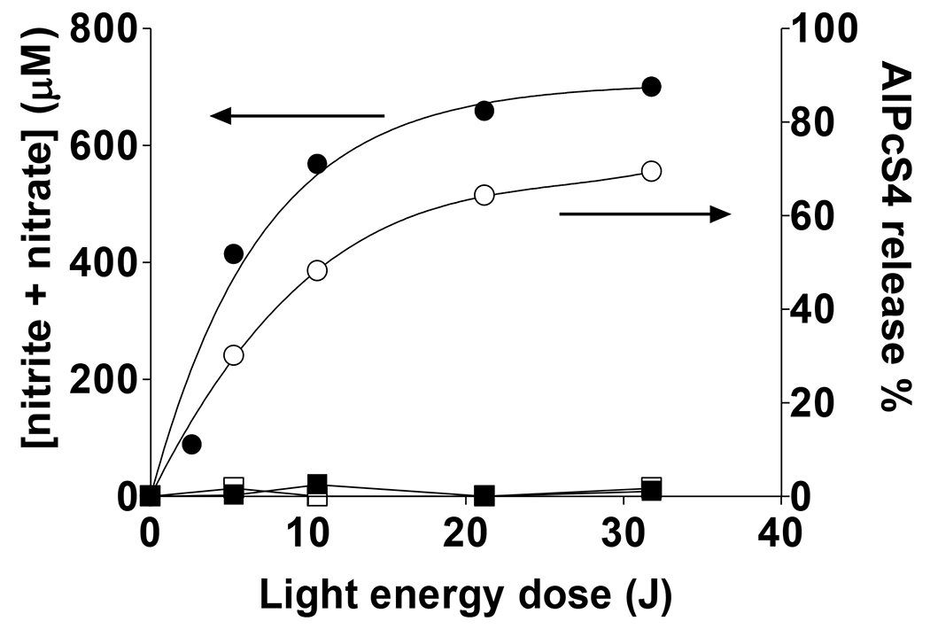
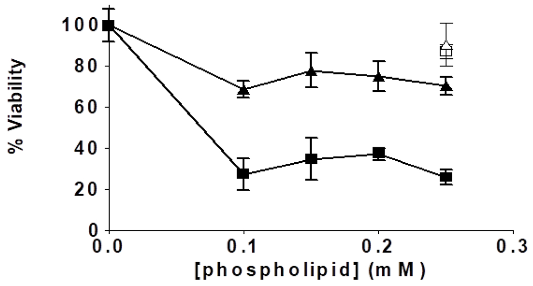
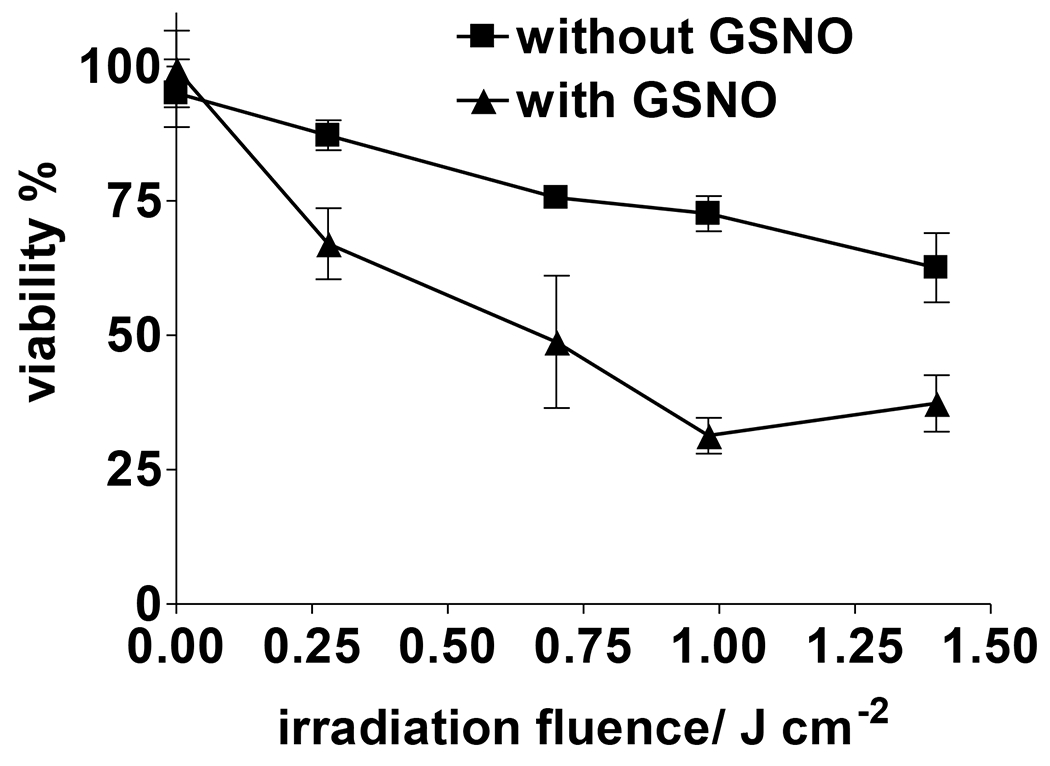
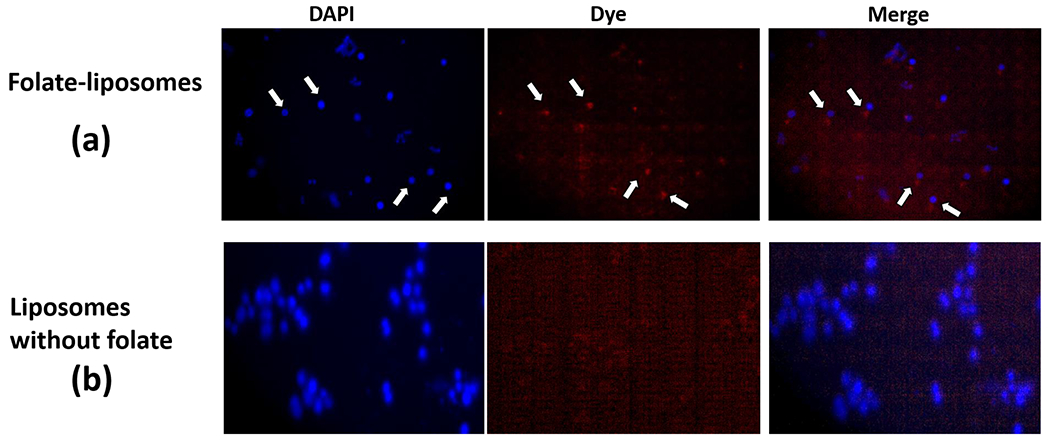
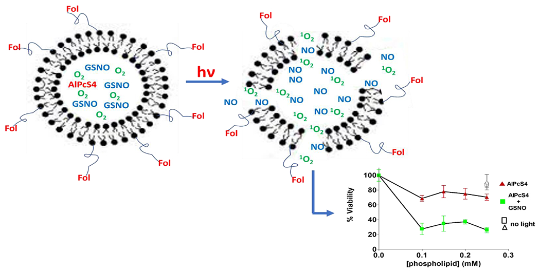
Near micromolar concentrations of nitric oxide (NO) induce tumor cells death. However, an appropriate NO load has to be delivered selectively to the tumor site in order to avoid NO loss and secondary NO-induced effects. The encapsulation of millimolar concentrations of a NO source and an appropriate trigger of NO release within phospatidylcholine-based liposomes should provide an efficient tool for the selective release of the needed NO payload. In this work we report the photosensitized generation of singlet oxygen and NO from folate-targeted PEGylated liposomes, containing AlPcS4 as the sensitizer and S-nitrosoglutathione (GSNO), in millimolar amounts, as the NO source. Amounts of singlet oxygen detected outside the liposome when using PEGylated liposomes are near 200 % larger when GSNO is present inside the liposomes as compared to its absence. These liposomes, conjugated to folate, were found to enhance the photosensitized cytotoxicity to A2780CP20 ovarian cancer cells as compared to liposomes containing the sensitizer but no GSNO (30 % as compared to 70 % cell viability) under the conditions of this work. Fluorescense of AlPcS4 was observed inside cells incubated with folate-conjugated liposomes but not with liposomes without folate. The photosensitized activity enhancement by GSNO increased when light fluence or liposome concentration were increased. The majority of ovarian cancer patients are initially diagnosed with disseminated intra-abdominal disease (stages III-IV) and have a 5-year survival of less than 20%. This work suggests a novel ovarian cancer nodules treatment via the use of tumor-targeted liposome nanoparticles with the capability of generating simultaneously reactive oxygen and nitrogen species upon illumination with near-infrared light.


Conflict of interest statement

Conflicts of Interest: The authors declare no conflict of interest. The funding sources had no role in study design, data collection, analysis and interpretation or in writing and submitting the manuscript.

Figures

Figure 1.
Figure 1.
Initial rates of photosensitized NO production upon irradiation of 60 mM GSNO and AlPcS4 at 670 nm in glycine buffer (pH 8.7) in the presence of 300 uM DTPA and 300 uM neocuproine. Samples were N2-saturated before starting irradiation. The radiation energy flux used was 70 mJ cm−2 s−1.
Figure 2.
Figure 2.
Particle size distribution of liposomes after 0 (white bars: (a) and (e)) or 10.6 J (colored bars) of irradiation at 670 nm. The liposome used in (a) – (d) is 6DOPC:4CHOL. The liposome used in (e) and (f) is 57.5DOPC:38.5CHOL:5.0PEG. All liposomes with the exception of that in (c) are air-saturated. Bulk concentrations of reagents before extrusion are 900 μM AlPcS4, 60 mM GSNO, 300 μM DETAPAC and 300 μM neocuproine in 20 mM glycine buffer at pH 8.7. (c) N2-saturated; (d) no GSNO was included. Phospholipid concentration was the same in all samples.
Figure 3.
Figure 3.
Nitrite + nitrate production (black symbols) and % AlPcS4 release (white symbols) from 6DOPC:4CHOL liposomes. Samples were irradiated (circles) or not irradiated (squares) at 670 nm. Non-irradiated samples were kept under dark during the time period used for irradiated samples. Bulk concentrations of reagents before liposome extrusion are 900 μM AlPcS4, 60 mM GSNO, 300 μM DETAPAC and 300 μM neocuproine in 20 mM glycine buffer at pH 8.7. Near 11 % of the nitrite + nitrate concentration corresponds to nitrate. NO to phospholipid (PL) mole ratios are shown in Table 2.
Figure 4.
Figure 4.
Western blot analysis of the FRα protein in A2780 and A2780CP20 cells. (A) Representative image of a Western blot analysis to detect the FRα levels. Western blots were performed with 50 μg of protein extracts. (B) Membranes were incubated with the secondary antibody, only. (C) Membrane was first incubated with a recombinant FRα protein an then incubated with the primary antibody followed by incubation with the secondary antibody.
Figure 5.
Figure 5.
Role of GSNO in the photosensitized cytotoxicity to A2780CP20 ovarian cancer cells after 1.4 J/cm2 of irradiation at 670 nm in the presence of different concentrations of 57.44DOPC:37.44CHOL:5DSPE(2000)PEG:0.13DSPE-PEG(5000)-Folate liposomes. Closed (or black) square and triangle symbols correspond to liposomes containing and not containing GSNO, respectively. Open (or white) symbols correspond to samples containing liposomes with 0.25 mM phospholipid with the same composition as those of the corresponding closed symbols and which were kept under dark during all the procedure. Bulk concentrations of reagents before liposome extrusion are 900 μM AlPcS4, 60 mM GSNO, 300 μM DETAPAC and 300 μM neocuproine in 20 mM glycine buffer at pH 8.7. Data presented are averages of 3 determinations ± SEM (P < 0.05 in two-tailed paired t-test comparing cell death with and without GSNO).
Figure 6.
Figure 6.
Role of light fluence and of GSNO presence in the photosensitized cytotoxicity to A2780CP20 ovarian cancer cells upon irradiation at 670 nm in the presence 57.44DOPC:37.44CHOL:5DSPE(2000)PEG:0.13DSPE-PEG(5000)-Folate liposomes at a constant PL concentration of 0.25 mM. Bulk concentrations of reagents before liposome extrusion are 900 μM AlPcS4, 60 mM GSNO, 300 μM DETAPAC and 300 μM neocuproine in 20 mM glycine buffer at pH 8.7. Data presented are averages of 3 determinations ± SEM (P < 0.05 in two-tailed paired t-test comparing cell death with and without GSNO).
Figure 7.
Figure 7.
Representative fluorescence emission intensity images after incubation of A2780CP20 cells with (a) 57.44DOPC:37.44CHOL:5.0PEG:0.13DSPE-PEG(5000)-Folate and (b) 57.5DOPC:37.5CHOL:5.0PEG liposomes, both containing AlPcS4 and GSNO. Procedures are described in MATERIALS AND METHODS.
Scheme 1.
Scheme 1.
Graphical representation of the results described in this work regarding liposome light activation of the liposomes used and the consequent A2780CP20 ovarian cancer cells viabilities.

Similar articles

Cited by

References

    1. Shen Z, Ma Q, Zhou X, Zhang G, Hao G, Sun Y et al. (2021) Strategies to improve photodynamic therapy efficacy by relieving the tumor hypoxia environment. NPG Asia Mater, 13:1–19.
    1. Di Nicola M, Williams BK Jr, Hua J, Bekerman VP, Mashayekhi A, Shields JA et al. (2021) Photodynamic Therapy (PDT) for Retinal Hemangioblastoma: Treatment Outcomes in 17 Consecutive Patients. Ophthalmol Retina,. - PubMed
    1. Rak J, Pouckova P, Benes J, Vetvicka D (2019) Drug delivery systems for phthalocyanines for photodynamic therapy. Anticancer Res 39:3323–39. - PubMed
    1. Shi Z, Meng X, Zhang K, Tang S, Zhang C, Yang Z et al. (2021) Engineering Structural Metal–Organic Framework for Hypoxia-Tolerant Type I Photodynamic Therapy against Hypoxic Cancer. ACS Mater Lett 3:781–9.
    1. Nwogu C, Kloc A, Attwood K, Bshara W, Durrani F, Pandey R (2021) Porfimer Sodium Versus PS785 for Photodynamic Therapy (PDT) of Lung Cancer Xenografts in Mice (A Novel Agent for Lung Cancer PDT). J Surg Res 263:245–50. - PubMed