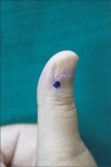
A Curious Case of Paroxysmal Painful Thumb: Glomus Tumor Re-visited
- PMID: 37035587
- PMCID: PMC10081463
- DOI: 10.4103/JCAS.JCAS_211_20
A Curious Case of Paroxysmal Painful Thumb: Glomus Tumor Re-visited
Abstract
Glomus tumor is a rare and benign vascular hamartoma originating from the neuro-myo-arterial apparatus of the glomus body in the reticular dermis. Most commonly, it presents as a solitary subungual lesion which is often painful and triggered by changes in temperature and pressure on the affected digit. However, multiple glomus tumors both digitally and elsewhere on the body which may or may not be painful are also known to occur. Due to its cryptic location and varied presentation, there is an invariable delay in the diagnosis and management of this condition. We report a case of glomus tumor in a 28-year-old woman presenting with paroxysmal painful lesion over her right thumb for 5 years.
Keywords: Glomus body; Love’s sign; ildreth’s sign; magnetic resonance imaging.
Copyright: © 2023 Journal of Cutaneous and Aesthetic Surgery.
Conflict of interest statement
There are no conflicts of interest.
Figures


Similar articles
-
Bilateral Solitary Glomus Tumour of Thumb: A Case Report.J Clin Diagn Res. 2017 May;11(5):RD04-RD06. doi: 10.7860/JCDR/2017/22374.9949. Epub 2017 May 1. J Clin Diagn Res. 2017. PMID: 28658864 Free PMC article.
-
Diagnosis and surgical approach in treating glomus tumor distal phalanx left middle finger: A case report.Int J Surg Case Rep. 2023 Jul;108:108426. doi: 10.1016/j.ijscr.2023.108426. Epub 2023 Jun 18. Int J Surg Case Rep. 2023. PMID: 37392587 Free PMC article.
-
A painful glomus tumor on the pulp of the distal phalanx.J Korean Neurosurg Soc. 2010 Aug;48(2):185-7. doi: 10.3340/jkns.2010.48.2.185. Epub 2010 Aug 31. J Korean Neurosurg Soc. 2010. PMID: 20856673 Free PMC article.
-
[Glomus tumors: anatomoclinical study of 14 cases with literature review].Ann Chir Plast Esthet. 2009 Feb;54(1):51-6. doi: 10.1016/j.anplas.2008.05.001. Epub 2008 Oct 19. Ann Chir Plast Esthet. 2009. PMID: 18938010 Review. French.
-
Large solitary glomus tumor of the wrist involving the radial artery.Am J Orthop (Belle Mead NJ). 2014 Dec;43(12):567-70. Am J Orthop (Belle Mead NJ). 2014. PMID: 25490012 Review.
References
-
- Lee JK, Kim TS, Kim DW, Han SH. Multiple glomus tumours in multidigit nail bed. Handchir Mikrochir Plast Chir. 2017;49:321–5. - PubMed
-
- Al-Qattan MM, Al-Namla A, Al-Thunayan A, Al-Subhi F, El-Shayeb AF. Magnetic resonance imaging in the diagnosis of glomus tumours of the hand. J Hand Surg Br. 2005;30:535–40. - PubMed
Publication types
LinkOut - more resources
Full Text Sources
