. 2023 Feb 28;14(14):3742-3751.
doi: 10.1039/d3sc00267e. eCollection 2023 Apr 5.

An abiotic, tetrameric, eight-helix bundle

Affiliations

An abiotic, tetrameric, eight-helix bundle

Friedericke S Menke et al. Chem Sci. .

Abstract

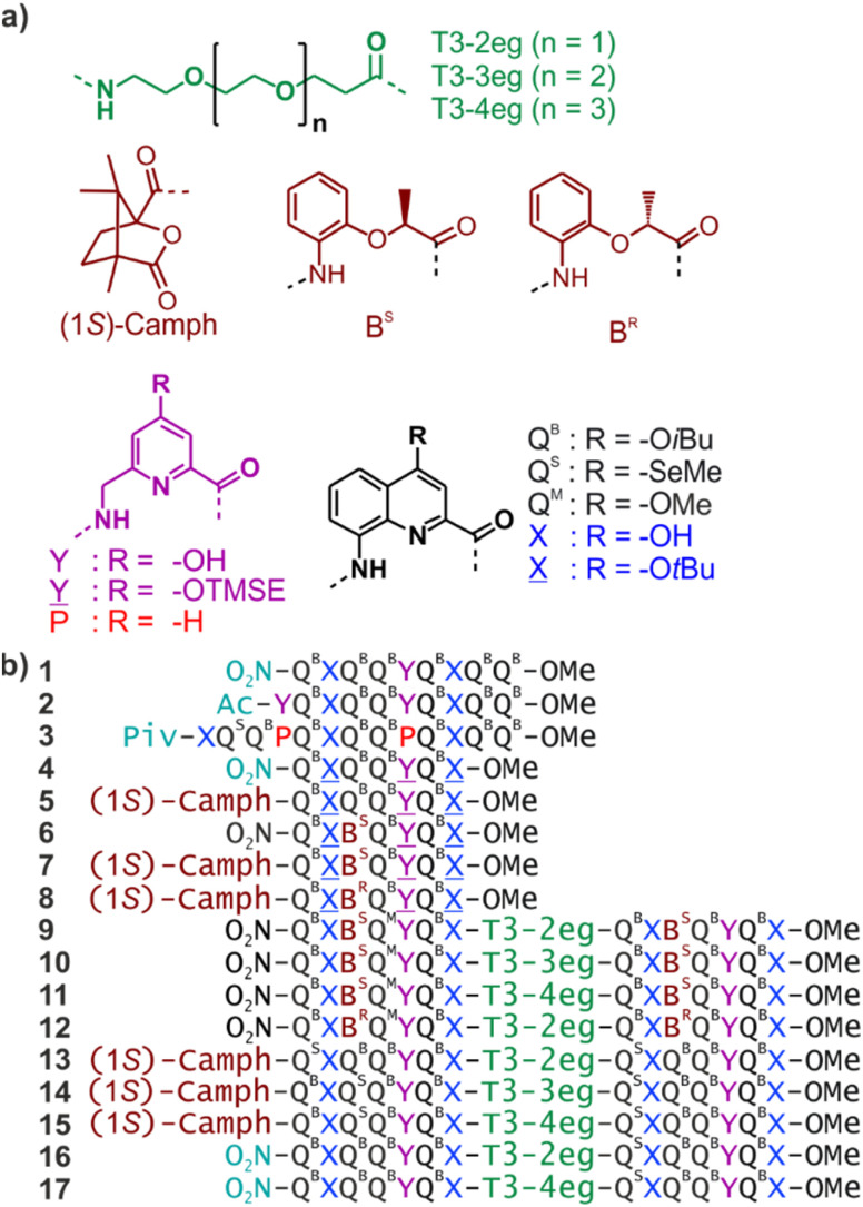
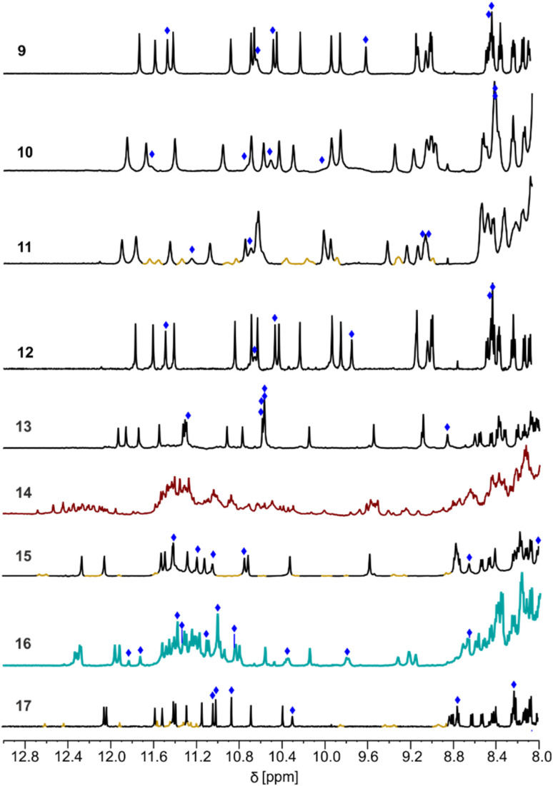
Four helically folded aromatic oligoamide sequences containing either a chiral monomer based on 2-(2-aminophenoxy)-propionic acid, an N-terminal (1H)-camphanyl group, or both, were synthesized. Spectroscopic solution investigations using 1H NMR and circular dichroism (CD) demonstrated that the 2-(2-aminophenoxy)-propionic acid unit biases helix handedness quantitatively in chloroform and dichloromethane. It even quantitatively overcomes an opposing effect of the camphanyl group and thus ensures reliable helix handedness control. A series of nine sequences composed of two helically folded aromatic oligoamide segments separated by a flexible linker based on a di-, tri- or tetraethylene glycol unit were then synthesized. In these sequences, helix handedness was controlled by means of an N-terminal (1H)-camphanyl group or a 2-(2-aminophenoxy)-propionic acid units in either both helical segments, or only in the N-terminal segment, or in none of the segments. The helical segments all displayed hydroxy and carbonyl groups at their surfaces as hydrogen bond donors and acceptors so as to promote helix-to-helix hydrogen bonding. NMR and CD spectroscopic studies showed that, in some cases, well-defined, stable, discrete abiotic helix-turn-helix tertiary folds form in organic solvents. Molecular modelling suggests that these correspond to structures in which the two helix axes are at an angle. In one case, the absence of handedness control resulted in a complex and large aggregate. A solid state structure obtained by single crystal X-ray diffraction analysis revealed a tetrameric assembly composed of eight helices with both right and left handedness arranged in three subdomains consisting of two hydrogen-bonded three-helix bundles and one two-helix-bundle. Several helix-to-helix hydrogen bonds were mediated by bridging water molecules. This structure constitutes an important milestone in the construction of abiotic protein-like architectures.


Conflict of interest statement

There are no conflicts to declare.

Figures

Fig. 1
Fig. 1. (a) Structures of di-, tri- and tetraethylene glycol T3 spacers, chiral (1S)-camph, BS and BR units, and Q, X, P and Y amino acid monomers. QB carries organic solubilizing side chains. QS was introduced in some sequences to assist crystallographic structure elucidation using the anomalous scattering of Se, though it turned out to be unneeded. QM is isosteric to QS. X̲ and Y̲ are the protected precursors of hydrogen-bonding monomers X and Y, respectively. TMSE = 2-trimethylsilylethyl. (b) Oligoamide foldamer sequences. Piv = pivaloyl. In sequences ending with an 8-nitro group, this group replaces the terminal amine.
Fig. 2
Fig. 2. (a) Cartoons representing identified assembly modes of aromatic helices displaying hydrogen bond acceptors and donors shown as red and yellow balls, respectively. The bottom left structure exists only when the helices are covalently linked. (b) Hydrogen-bonding patterns involving X and Y units as observed in helix-turn-helix tertiary structures in which sequences 1 or 2 are connected by a rigid linker. (c) Hydrogen-bonding patterns involving X and Y units as observed in a tilted dimer of 1. (d) Hydrogen-bonding patterns involving X and P units as observed in a PM (left) and in PP or MM (right) shifted dimers of 3. In (a–d), yellow and red circles around the hydrogen bond donors and acceptors correspond to the yellow cups and red knobs in (a) and Fig. 3, and to the yellow and red balls in Fig. 7.
Fig. 3
Fig. 3. Schematic representations of helix–helix hydrogen bonding when three hydrogen bond donors (yellow knobs) and three acceptors (red cups) are arranged in a sort of hexagon at the surface of a helix, as in 1. The hexagonal arrangement stems from two X units flanking a Y unit at the surface of a helix, with the hydrogen bond donor and acceptor being closer in X than in Y units (Fig. 2b). The helix face is represented as a plane, with the N- and C-termini shown as white and grey balls, respectively. Planes with a blue and red frame correspond to P and M helices, respectively. (a) “Open-book view” and stacked view of a hydrogen-bonded parallel head-to-head arrangement of two P helices and of the related clockwise and counterclockwise dimers. Hydrogen bonds are shown as dashed lines in the open-book view. In the stacked view, the top plane is transparent so that one can see the six hydrogen bonds (knobs into cups) and the plane behind. The parallel head-to-head arrangement has been observed only when the two helices are linked by a rigid turn unit (Fig. 2a and S1†). The PP clockwise tilted dimer of 1 has been characterized in the solid state. (b) Similar parallel arrangement, but this time head-to-tail, between a P and an M helix, as well as the clockwise and counterclockwise tilted dimers. The parallel arrangement has also been seen only when the helices are connected by a rigid linker. There is no evidence for the existence of PM tilted dimers, but models show plausible structures (see the ESI†).
Fig. 4
Fig. 4. Energy minimized model of a PP clockwise tilted helix-T3-2eg-helix arrangement. The sequence is analogous to that of 16 using Q units without side chains. (a) Space-filling representation with T3-2eg shown in green and the helices shown in blue. Amide carbonyl oxygen and hydroxy oxygen atoms involved in hydrogen bonding are shown in red. Hydroxy protons are shown in white. (b) Same view as in (a) showing only the outer rim of the backbone in tube representation. Hydrogen bond acceptors (carbonyl oxygen atoms) and donors (hydroxy protons) are shown as balls. (c) Same representation as in (b) but a different view showing the pseudo-symmetry of the structure (with the exclusion of the T3-2eg linker) and approximately hexagonal arrangement of the hydrogen bond donors and acceptors.
Fig. 5
Fig. 5. Part of the 500 MHz 1H NMR spectra of sequences 9–17 at 2.3 mM in CDCl3 at 25 °C showing the amide and hydrogen-bonded OH proton resonances. Signals assigned to OH protons are marked with a blue diamond. A few OH protons could not be unambiguously assigned due to signal overlap. For 11, 15 and 17, signals assigned to different aggregates are shown in different colors.
Fig. 6
Fig. 6. Structure of 164 in the solid state. (a) Top view and side view of the tetramer shown in a space-filling representation. The four molecules are shown in blue, red, gold and green. (b) Side view and front view of the tetramer in tube representation. The same colors are used as in (a). The inversion center is indicated by a black dot (i). Water molecules hydrogen bonded at helix–helix interfaces are shown in space-filling representation. Side chains and other included solvent molecules are omitted for clarity.
Fig. 7
Fig. 7. (a) Side-view of the crystal structure of 164 in tube representation indicating the three different domains. The four molecules are shown in brown (A), light blue (B) red (B′) and dark blue (A′). The oxygen atoms at position 4 of X and Y units involved in hydrogen bonding are shown as yellow balls. Those not involved in hydrogen bonding are shown as purple balls. The oxygen atoms of amide carbonyl groups involved in hydrogen bonding are shown as red balls. Bridging water molecules are shown as green balls. Hydroxy protons and carbonyl oxygen atoms of the hydrogen-bonding arrays are shown as yellow and red balls, respectively. Hydrogen atoms and side chains are not shown for clarity. (b) Top view of a three-helix domain with the same representation as in (a). (c) Side view of the central two-helix domain with the same representation as in (a). Arrows indicate the N → C orientation of the sequences. Green circles highlight areas of interest that are shown in (d). (d) Water molecule hydrogen bonded simultaneously to three different helices, including one T3-2eg linker and the carbonyl at position 4 of the C-terminal X unit of molecule B, found as its (1H)-4-quinolinone tautomer.

Similar articles

Cited by

References

    1. Guichard G. Huc I. Chem. Commun. 2011;47:5933. doi: 10.1039/C1CC11137J. - DOI - PubMed
    1. Gellman S. H. Acc. Chem. Res. 1998;31:173–180. doi: 10.1021/ar960298r. - DOI
    1. Goodman C. M. Choi S. Shandler S. DeGrado W. F. Nat. Chem. Biol. 2007;3:252–262. doi: 10.1038/nchembio876. - DOI - PMC - PubMed
    1. Nair R. V. Vijayadas K. N. Roy A. Sanjayan G. J. Eur. J. Org. Chem. 2014;2014:7763–7780. doi: 10.1002/ejoc.201402877. - DOI
    1. Saraogi I. Hamilton A. D. Chem. Soc. Rev. 2009;38:1726. doi: 10.1039/B819597H. - DOI - PubMed