Lipid oxidation controls peptide self-assembly near membranes through a surface attraction mechanism
- PMID: 37035708
- PMCID: PMC10074436
- DOI: 10.1039/d3sc00159h
Lipid oxidation controls peptide self-assembly near membranes through a surface attraction mechanism
Abstract
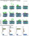
The self-assembly of peptides into supramolecular structures has been linked to neurodegenerative diseases but has also been observed in functional roles. Peptides are physiologically exposed to crowded environments of biomacromolecules, and particularly cellular membrane lipids. Previous research has shown that membranes can both accelerate and inhibit peptide self-assembly. Here, we studied the impact of membrane models that mimic cellular oxidative stress and compared this to mammalian and bacterial membranes. Using molecular dynamics simulations and experiments, we propose a model that explains how changes in peptide-membrane binding, electrostatics, and peptide secondary structure stabilization determine the nature of peptide self-assembly. We explored the influence of zwitterionic (POPC), anionic (POPG) and oxidized (PazePC) phospholipids, as well as cholesterol, and mixtures thereof, on the self-assembly kinetics of the amyloid β (1-40) peptide (Aβ40), linked to Alzheimer's disease, and the amyloid-forming antimicrobial peptide uperin 3.5 (U3.5). We show that the presence of an oxidized lipid had similar effects on peptide self-assembly as the bacterial mimetic membrane. While Aβ40 fibril formation was accelerated, U3.5 aggregation was inhibited by the same lipids at the same peptide-to-lipid ratio. We attribute these findings and peptide-specific effects to differences in peptide-membrane adsorption with U3.5 being more strongly bound to the membrane surface and stabilized in an α-helical conformation compared to Aβ40. Different peptide-to-lipid ratios resulted in different effects. We found that electrostatic interactions are a primary driving force for peptide-membrane interaction, enabling us to propose a model for predicting how cellular changes might impact peptide self-assembly in vivo.
This journal is © The Royal Society of Chemistry.
Conflict of interest statement
There are no conflicts to declare.
Figures


Similar articles
-
Origin of Secondary Structure Transitions and Peptide Self-Assembly Propensity in Trifluoroethanol-Water Mixtures.J Phys Chem B. 2024 Aug 15;128(32):7736-7749. doi: 10.1021/acs.jpcb.4c02594. Epub 2024 Aug 1. J Phys Chem B. 2024. PMID: 39088441
-
Molecular Understanding of Aβ-hIAPP Cross-Seeding Assemblies on Lipid Membranes.ACS Chem Neurosci. 2017 Mar 15;8(3):524-537. doi: 10.1021/acschemneuro.6b00247. Epub 2016 Dec 12. ACS Chem Neurosci. 2017. PMID: 27936589
-
Secondary Structure Transitions for a Family of Amyloidogenic, Antimicrobial Uperin 3 Peptides in Contact with Sodium Dodecyl Sulfate.Chempluschem. 2022 Jan;87(1):e202100408. doi: 10.1002/cplu.202100408. Chempluschem. 2022. PMID: 35032115
-
Peptide Self-Assembly into Amyloid Fibrils at Hard and Soft Interfaces-From Corona Formation to Membrane Activity.Macromol Biosci. 2023 Jun;23(6):e2200576. doi: 10.1002/mabi.202200576. Epub 2023 Mar 10. Macromol Biosci. 2023. PMID: 36810963 Review.
-
Peptide-lipid interactions of the beta-hairpin antimicrobial peptide tachyplesin and its linear derivatives from solid-state NMR.Biochim Biophys Acta. 2006 Sep;1758(9):1285-91. doi: 10.1016/j.bbamem.2006.03.016. Epub 2006 Apr 5. Biochim Biophys Acta. 2006. PMID: 16678119 Review.
Cited by
-
Nonequilibrium Self-Assembly Control by the Stochastic Landscape Method.J Chem Inf Model. 2025 Apr 28;65(8):4067-4080. doi: 10.1021/acs.jcim.4c02366. Epub 2025 Apr 8. J Chem Inf Model. 2025. PMID: 40198896 Free PMC article.
-
Carbene formation as a mechanism for efficient intracellular uptake of cationic antimicrobial carbon acid polymers.Nat Commun. 2025 Jul 12;16(1):6460. doi: 10.1038/s41467-025-61724-y. Nat Commun. 2025. PMID: 40652000 Free PMC article.
-
Lipid-polymer hybrid-vesicles interrupt nucleation of amyloid fibrillation.RSC Chem Biol. 2024 Oct 24;5(12):1248-1258. doi: 10.1039/d4cb00217b. eCollection 2024 Nov 27. RSC Chem Biol. 2024. PMID: 39569389 Free PMC article.
-
Bio-Inspired Self-Assembly of Enzyme-Micelle Systems for Semi-Artificial Photosynthesis.Angew Chem Int Ed Engl. 2025 Apr 25;64(18):e202424222. doi: 10.1002/anie.202424222. Epub 2025 Mar 3. Angew Chem Int Ed Engl. 2025. PMID: 39951445 Free PMC article.
References
LinkOut - more resources
Full Text Sources

