

Clinical and genetic analysis in Chinese families with synpolydactyly, and cellular localization of HOXD13 with different length of polyalanine tract
- PMID: 37035736
- PMCID: PMC10073534
- DOI: 10.3389/fgene.2023.1105046
Clinical and genetic analysis in Chinese families with synpolydactyly, and cellular localization of HOXD13 with different length of polyalanine tract
Abstract
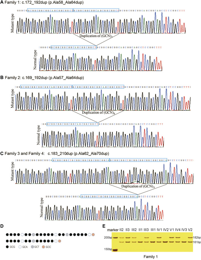
Synpolydactyly (SPD) is caused by mutations in the transcription factor gene HOXD13. Such mutations include polyalanine expansion (PAE), but further study is required for the phenotypic spectrum characteristics of HOXD13 PAE. We investigated four unrelated Chinese families with significant limb malformations. Three PAEs were found in the HOXD13 polyalanine coding region: c.172_192dup (p.Ala58_Ala64dup) in Family 1, c.169_192dup (p.Ala57_Ala64dup) in Family 2, and c.183_210dup (p.Ala62_Ala70dup) in Family 3 and Family 4. Interestingly, we identified a new manifestation of preaxial polydactyly in both hands in a pediatric patient with an expansion of seven alanines, a phenotype not previously noted in SPD patients. Comparing with the wild-type cells and mutant cells with polyalanine contractions (PACs), the HOXD13 protein with a PAE of nine-alanine or more was difficult to enter the nucleus, and easy to form inclusion bodies in the cytoplasm, and with the increase of PAE, the more inclusion bodies were formed. This study not only expanded the phenotypic spectrum of SPD, but also enriched our understanding of its pathogenic mechanisms.
Keywords: HOXD13; polyalanine expansion; preaxial polydactyly; synpolydactyly; variant.
Copyright © 2023 Chen, Zhao, Xu, Cao, Li, Zhang and Zhao.
Conflict of interest statement
The authors declare that the research was conducted in the absence of any commercial or financial relationships that could be construed as a potential conflict of interest.
Figures


Similar articles
-
New insight into the development of synpolydactyly caused by expansion of HOXD13 polyalanine based on weighted gene co-expression network analysis.BMC Med Genomics. 2024 Oct 29;17(1):259. doi: 10.1186/s12920-024-01974-9. BMC Med Genomics. 2024. PMID: 39472920 Free PMC article.
-
A Novel Missense Variant of HOXD13 Caused Atypical Synpolydactyly by Impairing the Downstream Gene Expression and Literature Review for Genotype-Phenotype Correlations.Front Genet. 2021 Oct 27;12:731278. doi: 10.3389/fgene.2021.731278. eCollection 2021. Front Genet. 2021. PMID: 34777468 Free PMC article.
-
[HOXD13 polyalanine tract expansion in synpolydactyly: mutation detection and prenatal diagnosis in a large Chinese family].Zhonghua Yi Xue Yi Chuan Xue Za Zhi. 2005 Feb;22(1):5-9. Zhonghua Yi Xue Yi Chuan Xue Za Zhi. 2005. PMID: 15696469 Chinese.
-
HOXD13-associated synpolydactyly: Extending and validating the genotypic and phenotypic spectrum with 38 new and 49 published families.Genet Med. 2023 Nov;25(11):100928. doi: 10.1016/j.gim.2023.100928. Epub 2023 Jul 7. Genet Med. 2023. PMID: 37427568 Review.
-
Limb skeletal malformations - what the HOX is going on?Eur J Med Genet. 2012 Jan;55(1):1-7. doi: 10.1016/j.ejmg.2011.06.003. Epub 2011 Jul 2. Eur J Med Genet. 2012. PMID: 21782042 Review.
Cited by
-
New insight into the development of synpolydactyly caused by expansion of HOXD13 polyalanine based on weighted gene co-expression network analysis.BMC Med Genomics. 2024 Oct 29;17(1):259. doi: 10.1186/s12920-024-01974-9. BMC Med Genomics. 2024. PMID: 39472920 Free PMC article.
References
LinkOut - more resources
Full Text Sources