Review
. 2023 Mar 22;5(1):20220054.
doi: 10.1259/bjro.20220054. eCollection 2023.

Dual-energy CT for gastrointestinal bleeding

Affiliations
Review

Dual-energy CT for gastrointestinal bleeding

Miyuki Okamura-Kawasaki et al. BJR Open. .

Abstract

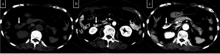
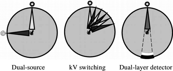
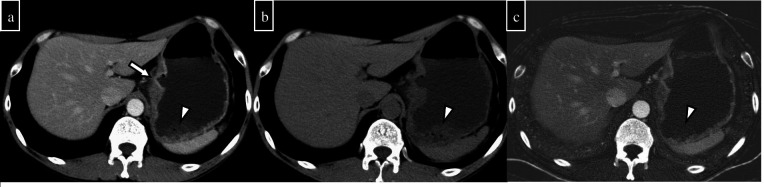
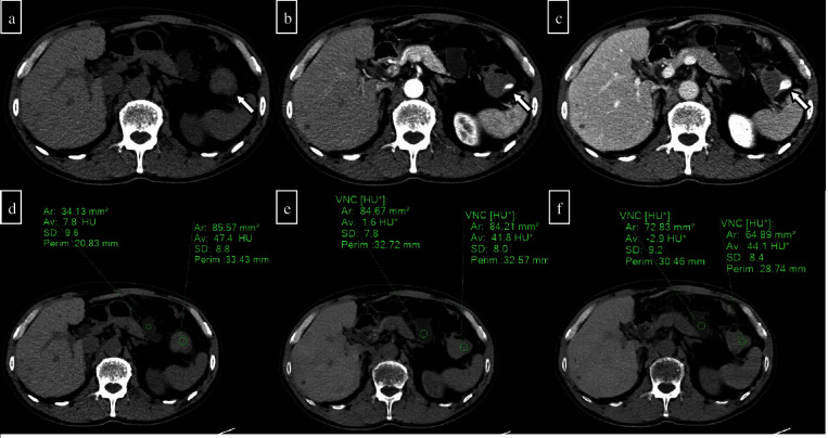
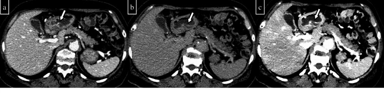
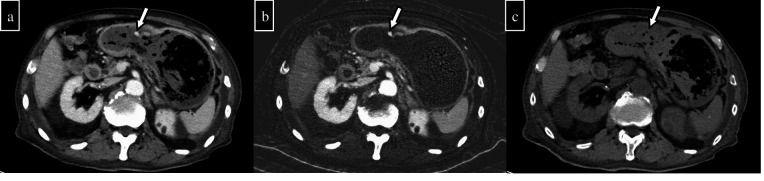
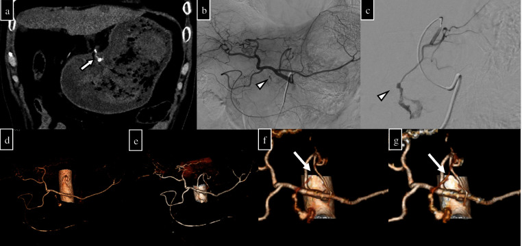
Dual-energy computed tomography (DECT) can be used for various types of analyses, including iodine quantification, and its usefulness in diagnosing gastrointestinal diseases has been reported. This pictorial review describes the use of DECT in the diagnosis of gastrointestinal bleeding. Virtual non-contrast computed tomography (CT) is available in DECT and can be used as a substitute for pre-contrast CT in the case of gastrointestinal haemorrhage. The omission of pre-contrast CT can reduce radiation exposure by approximately 30%. A low-keV virtual monochromatic X-ray image (VMI) can increase the contrast of iodine, and iodine maps can provide better visibility of extravasation. These analytical images can provide a diagnosis with a high degree of confidence. In addition, the low-keV VMI clearly illustrates the vascular structure, which may be useful for improving the visibility of vascular lesions and for confirming the arterial anatomy before embolisation. Considering that these analytical images are created on the basis of contrast-enhanced CT, the positional information of organs is entirely identical, thus allowing the comparison of images regardless of intestinal peristalsis or body motion. In conclusion, the analytical images of DECT can solve the problems of conventional protocols, and DECT is considered useful in the imaging diagnosis of gastrointestinal bleeding.


Figures

Figure 1.
Figure 1.
An 87-year-old woman had progressive anaemia and underwent pre-contrast and post-contrast two-phase (arterial and delayed phase) CT. (a) On the pre-contrast CT, it is difficult to determine whether the duodenal contents (arrow) are haematomas. (b) The arterial phase shows extravasation into the duodenal lumen. (c) Extravasation increases in the delayed phase. On the basis of these findings, duodenal active bleeding is suspected.
Figure 2.
Figure 2.
Various methods of DECT. DECT with dual-source, kV switching, and dual-layer detector are simulated from left to right. DECT, dual-energy computed tomography.
Figure 3.
Figure 3.
A 52-year-old man has a history of gastric ulcer bleeding. Owing to haematogenous vomiting, single-phase CT was performed. (a) Conventional CT shows an ulcer in the gastric lesser curvature (arrow). Gastric contents show slightly high density on conventional CT (arrowheads). (b) VNC shows a slightly high density of gastric contents (arrowheads). (c) The iodine map shows no apparent contrast agent content in the gastric contents. Haematoma is suspected rather than active bleeding. No obvious bleeding was observed on the endoscopy. VNC, virtual non-contrast CT.
Figure 4.
Figure 4.
A 70-year-old man presented to the clinic for bleeding after the endoscopic mucosal resection of a colonic polyp and underwent pre- and post-contrast two-phase CT (arterial and delayed phases). (a) Pre-contrast CT showed a relatively high density of contents in the splenic flexure of the colon, thus suggesting a haematoma (arrow). (b) The arterial phase shows extravasation in the splenic flexure. (c) Extravasation is increasing in the delayed phase. From the above, active haemorrhage after endoscopic mucosal resection was suspected. (d) Pre-contrast CT measurement shows a 47.4 HU in the splenic flexure lumen and a 7.8 HU in the small intestine lumen in the area nearby. (e) In the VNC created from arterial phase images, the densities of the splenic flexure and the small intestine in the nearby area are 41.8 and 1.6 HU, respectively. (f) In the VNC created from the delayed phase image, the densities in the splenic flexure and the small intestine in the nearby area are 44.1 and -2.9 HU, respectively. There is no significant difference that would quantitatively affect the evaluation. VNC, virtual non-contrast CT.
Figure 5.
Figure 5.
A 62-year-old woman presented with complaints of black stools and hypotension to the hospital. A contrast-enhanced two-phase (portal venous phase and delayed phase six minutes later) CT was performed to assess the cause of bleeding. (a) In the portal venous phase, a punctate high-density area was found near the gastric pylorus (arrow). (b) A punctate high-density area is seen near the gastric pylorus in the delayed phase, and the high-density area is slightly enlarged compared with that in the portal venous phase. This is considered an active haemorrhage. (c) The VMI (40 keV) of the delayed phase shows a clear spread of contrast agent compared with the original image. VMI, virtual monochromatic image.
Figure 6.
Figure 6.
An 81-year-old man was hospitalised for heart failure because of atrial fibrillation. A single-phase contrast-enhanced CT was performed because of sudden haematochezia. (a) Conventional CT shows a high-density area in the stomach (arrow). (b) The iodine map shows high density in the same area and low density in all other gastric contents. (c) VNC shows a decrease in the density of the high-density area in the stomach on conventional CT. From (b) and (c), the presence of active bleeding in the stomach can be diagnosed. VNC, virtual non-contrast CT.
Figure 7.
Figure 7.
A 63-year-old man. Eight days after omental plugging for gastric perforation, he developed progressive anaemia and hypotension. Emergency endoscopy was performed, and a large amount of blood was found in the stomach. Given that endoscopic haemostasis was difficult, embolisation was decided, and pre- and post-contrast CT (arterial and delayed phases) were performed to identify the bleeding spot. (a) In the arterial phase (coronal section), an ulcer on the gastric kyphosis and a hyperintense area near the ulcer were observed, and extravasation was considered (arrow). (b) Abdominal arteriography and (c) left gastric arteriography show extravasation from the peripheral left gastric artery branch (arrowhead). (d) and (e) Vascular VR images created by conventional CT of the arterial phase and VMI (40 keV) images of the arterial phase with bone removal; no responsible vessels are seen in either case. (f) and (g) VR images created after extraction of the left gastric artery vessels using conventional CT of the arterial phase and VMI (40 keV) images of the arterial phase; in both images, the responsible branch is indicated (dashed arrows). The branches are more clearly depicted in the VR images created using VMI (40 keV) in both methods. VR, volume rendering; VMI, virtual monochromatic image.

Similar articles

Cited by

References

    1. Wortman JR, Landman W, Fulwadhva UP, Viscomi SG, Sodickson AD. CT angiography for acute gastrointestinal bleeding: what the radiologist needs to know. Br J Radiol 2017; 90: 20170076. doi: 10.1259/bjr.20170076 - DOI - PMC - PubMed
    1. Trabzonlu TA, Mozaffary A, Kim D, Yaghmai V. Dual-Energy CT evaluation of gastrointestinal bleeding. Abdom Radiol (NY) 2020; 45: 1–14. doi: 10.1007/s00261-019-02226-6 - DOI - PubMed
    1. Murray N, Darras KE, Walstra FE, Mohammed MF, McLaughlin PD, Nicolaou S. Dual-Energy CT in evaluation of the acute abdomen. Radiographics 2019; 39: 264–86. doi: 10.1148/rg.2019180087 - DOI - PubMed
    1. Wells ML, Hansel SL, Bruining DH, Fletcher JG, Froemming AT, Barlow JM, et al. . Ct for evaluation of acute gastrointestinal bleeding. Radiographics 2018; 38: 1089–1107. doi: 10.1148/rg.2018170138 - DOI - PubMed
    1. Mohammadinejad P, Kwapisz L, Fidler JL, Sheedy SP, Heiken JP, Khandelwal A, et al. . The utility of a dual-phase, dual-energy CT protocol in patients presenting with overt gastrointestinal bleeding. Acta Radiol Open 2021; 10: 20584601211030656. doi: 10.1177/20584601211030658 - DOI - PMC - PubMed